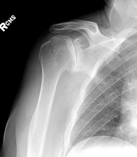
This patient presented with a chronic anterior dislocation of the humeral head after a proximal humerus fracture, the humeral head was malunited in the subscapularis recess. Osteotomy of the humeral head was required to remove the bone from the anterior glenoid. The fracture was addressed with hemiarthroplasty and repair of the tuberosities. The use of a stem that provides a "window" for proximal bone grafting, the removal of the cement circumferentially from the proximal stem during implantation, and the stable suture repair of the tuberosities with Fiberwire provided a good outcome with union of the tuberosities. The anterior inferior subluxation of the humeral head persisted for 6 months but eventually it was resolved. Contrary to the belief that this is the result of a "fracture hematoma" it seems that this finding is associated with muscle atony or contusion of the rotator cuff or axillary nerve neuropraxia. X-rays are shown below.
Tuesday, October 31, 2017
Monday, October 30, 2017
Locked proximal humerus fracture dislocation
Fracture locked dislocations of the proximal humerus are challenging injuries due to the energy of the trauma and the instability that is encountered during surgery. Small bony bankart lesions as in this case do not need to be fixed. The progressive development of stiffness usually balances the instability. If the fracture fixation is combined with labrum or bankart repair then there is a concern for development of significant stiffness. The following x-rays demonstrate a patient who was treated with ORIF of the humerus fracture and no repair of the small bony bankart lesion. She regained active elevation of her shoulder to 160 degrees without instability.
Friday, September 15, 2017
Post-traumatic stiffness and malunion after supracondylar humerus fracture in an adolescent patient
The following case illustrates a high degree of post-traumatic complexity due to the malunion of the distal humerus and osteophyte formation with bony block in motion. This was the sequela of a supracondylar humerus fracture on a patient who was close to skeletal maturity. Flexion extension was from 45 to 90 degrees. An attempt for arthroscopic osteoplasty was performed which was unsuccessful because there was no joint space. Conversion to open surgery was chosen. According to the technique described by Hill Hastings, MD we performed osteoplasty of the elbow. Through a medial and lateral elbow approach osteoplasty of the humerus was performed, resection of osteophytes from the anterior compartment, anterior capsular release and release of the posterior and transverse band of the medial collateral ligament. Flexion improved to 110 degrees extension to 20 degrees. The ulnar nerve was transposed.
After the osteoplasty range of motion improved significantly, especially in elbow flexion. The terminal extension was still compromised with a loss of 30 degrees of terminal extension.
Terrible triad injury. The importance of the LCL complex
Terrible triad injuries are high energy traumatic events to the elbow which are associated with significant soft tissue trauma. The following case illustrates an obese patient who suffered such an injury. The elbow was severely unstable while the weight of the arm was contributing to the instability as well. During surgery the following "inside to outside" algorithm for fixation is used. Fixation begins with the deep structures and we move superficially through a lateral approach. The sequence is the following;
1. Assessment of the coronoid fracture - if large fragment then fixation with sutures or with a screw is indicated
2. Radial head replacement
3. Primary repair of the lateral collateral ligament complex using bone tunnels at the humerus and no anchors. We avoid use of anchors that increase the risk of infection
If the elbow is stable after following the above 3 steps then repair of the medial collateral ligament complex is not indicated. In this case the MCL was ruptured as well however the elbow was stable after the completion of step 3 as illustrated above.
If the elbow is unstable after completion of the above 3 steps then the MCL is repaired. If still unstable then a static external fixator is applied for 6 weeks.
Key points for success of treatment are (a) avoidance of overstuffing of the joint by undersizing the prosthetic radial heads (b) stable fixation of the lateral collateral ligament in the acute setting with a Krackow suturing method. A common mistake is use of large radial prosthetic head "for better stability". Large heads increase the change of subluxation, pain and stiffness. The illustration below indicates the importance of anatomic stable fixation of the LCL using the Krackow method.
trauma films
first reduction was unsuccessful due to inadequate flexion of the elbow and weight of the arm.
Second attempt was successful in close reduction
Undersized the radial head implant is important. One method of assessing potential overstuffing of the joint is the following:
"Incongruity of the medial ulnohumeral joint which becomes apparent radiographically only after overlengthening of the radius by ≥6 mm. Intraoperative visualization of a gap in the lateral ulnohumeral joint is a reliable indicator of overlengthening following the insertion of a radial head prosthesis". We also use the congruency of the proximal radioulnar joint surface line as an indicator of appropriate height of the prosthesis. No step off should be seen.
Sprengel Deformity in adulthood -
This is a complex congenital condition which most of the time is associated with other syndromes or congenital defects. An alternative term used to describe this condition is scapula elevata. Patients may have a cosmetic deformity with a "high riding" scapula which is "hypomobile" and there is some degree of scapula winging. In severe forms of the disease there is a omovertebra bone with joint like connection between the cervical spine and the medial border of the scapula.
There are authors who have described good results after surgical intervention when the condition is diagnosed before the age of 6 . The main problem in this clinical condition is restricted range of shoulder motion. Most of the time forward elevation and abduction is limited, due to loss of the scapular motion or scapulohumeral rhythm as described by Codman in the 1930s.
When the loss of motion is minimal then surgery is usually contra-indicated. Associated syndromes such as Klippel Feil syndrome or other syndromes may compromise surgical care of this condition.
The following case illustrates a patient who present with the condition in adulthood and had no treatment before. The fused upper ribs and deformed thoracic cage indicate in this situation the failure of the scapula to decent during development. Associated hypoplasia of the affected scapula is not uncommon.
The forward elevation of the shoulder in the following case was around 100 degrees and for that reason no surgery was offered.
Wednesday, August 16, 2017
The stiff arthritic shoulder with flattened posteriorly decentered humeral head. Tips for a successful anatomic arthroplasty.
Posterior subluxation of the humeral head in primary osteoarthritis of the shoulder is common. It is usually associated with a B2 Walch glenoid deformity. When there is associated flattening of the humeral head, as seen in this case below, usually there is significant limitation is motion and stiffness.
When planning the total shoulder anatomic replacement, it is important not to proceed with an aggressive humeral head cut. Significant decrease of the volume of the head due to an aggressive humeral head cut will help with glenoid exposure BUT it will make the posterior instability worse. While some surgeons use posterior augmented glenoid components, I prefer to address the instability with soft tissue balancing, conservative humeral head cut, eccentric humeral head - dialing eccentricity anteriorly - and possible closure of the rotator cuff interval at the end of the surgery.
Below you will find xrays of a case that illustrates a clinical situation where such approach had to be used.


