Tuesday, May 20, 2014

Intramedullary nailing of bilateral pathologic humerus fractures

This a female patient in her seventies who presented in the office with arm pain. She has been diagnosed with multiple myeloma (most common bone tumor for that age group). She has lytic bone lesions to bilateral humeri as demonstrated in the images below with a pathologic humerus fracture on the left. On the left side there is a lytic lesion at the proximal metaphysis and a pathologic fracture at the distal metaphysis of the bone.


As demonstrated by these preoperative films there is an impending pathologic fracture on the R at the proximal metaphysis and a pathologic fracture on the left at the distal metaphysis.

The patient had no contraindications to surgery and for this reason the fractures were stabilized with antegrade humeral nailing. At one year she is pain free to bilateral arms. Xrays are shown below.


We prefer to use IM nailing for pathologic humerus fracture, however there is a risk of shoulder pain after placement of the nails. For this reason this implant is avoided in the young active healthy individuals. In addition, placement of the distal locking screw requires good surgical exposure and technique to avoid injury to the radial nerve or nerve structures at the distal aspect of the humerus.

Sunday, May 18, 2014

How to ream a biconcave glenoid and when to use an eccentric humeral head in total shoulder replacement

Biconcave glenoids are usually associated with slight or significant subluxation of the humeral head with an associated wear of the posterior aspect of the glenoid. The glenoid is dysplastic is this cases and the wear is usually seen at the posterior superior aspect of the glenoid. The following case of shoulder osteoarthritis with a biconcave glenoid was approached with reaming of the glenoid without correction of the deformity and with placement of an eccentric head with the eccentricity anteriorly to compensate for the posterior subluxation of the humeral head. Preoperative and postoperative xrays are shown below.
CLICK ON IMAGE FOR LARGER VERSION. Axillary view demonstrates the biconcave glenoid

CLICK ON IMAGE FOR LARGER VERSION. Grashey view demonstrates the glenohumeral arthritis 

Postoperative images below demonstrate the eccentric humeral head used for this case to avoid drop back of the humeral head during forward elevation of the shoulder. If there is still drop back after the placement of the trial eccentric head then rotator cuff interval closure is necessary.
CLICK ON IMAGE FOR LARGER VERSION. Axillary view demonstrates the eccentric humeral head

CLICK ON IMAGE FOR LARGER VERSION. Grashey view shows the position of the implants

There are two ways to ream a biconcave arthritic glenoid. One way requires no correction of the deformity and less bone removal and the other requires correction of the deformity and more bone removal. Generally, the reaming should not be done beyond the subchondral plate and preservation of bone stock at the glenoid is critical in the stability and longevity of the glenoid component. The more bone is removed the less cortical bone is left at the implant - glenoid bone interface to support the all polyethelene glenoid component. Implanted glenoids on cancellous bone run the risk of loosening in the short term. The drawings below demonstrate the two different methods of reaming a biconcave glenoid in total shoulder arthroplasty.
REAMING WITHOUT correction of the deformity. (A) a biconcave glenoid is depicted. (B) First with the use of a curette the biconcavity is eliminated and the biconcave glenoid is converted to a concave glenoid (C) the reaming is performed and minimal bone is removed until the reamer "sits" on the glenoid.

REAMING WITH correction of the deformity. (A) a biconcave glenoid is depicted (B) The reamer is levered more anteriorly to correct the deformity and convert the biconcave glenoid to a concave glenoid (C) the reaming is performed and MORE bone is removed compared to the previous example until the reamer "sits" on the glenoid

Comparing the two methods it is obvious that without correction of the deformity (A) there is no significant bone removal. With correction of the deformity (B) there is MORE bone removed compared to method (A).

(A) No correction of the deformity does not correct the posterior  subluxation of the humeral head (B) Correction of the glenoid deformity does correct to a certain degree the posterior subluxation of the humeral head. Notice the distance of the plane of the scapula relative to the center of the humeral head

(A) Placement of an eccentric head can compensate for the posteriorly subluxated humeral head (B) In cases where the glenoid deformity is corrected a concentric humeral head may or may not provide stability and correct posterior subluxation of the humeral head.


Saturday, May 17, 2014

Installing the World's First Electronic Spine. From The Washington Post

Ohio surgeons hope chip in man’s brain lets him control paralyzed hand with thoughts


(Courtesy of The Ohio State University Wexner Medical Center) - Neurosurgeons Milind Deogaonkar, left, and Ali Rezai, right, of the Ohio State University Wexner Medical Center perform brain surgery to implant the Neurobridge technology in a 22-year-old partially paralyzed man.


COLUMBUS, Ohio — Chad Bouton snapped awake at 5 a.m. He skipped coffee, threw some Clif Bars and water in a bag, and left his wife and children and robot at home. He ordered three Sausage McMuffins from a drive-up window. He steered toward the hospital. He entered the operating room at 6:15 a.m.
Bouton is a research engineer, not a doctor, and he worked with an engineer’s precision. By 7 a.m., he and his team had set everything up in the operating room and tested it — the computer with the secret algorithm, the uplink cable, the Wand. They were ready to go, right on time. As they had been told to expect, the patient was late. So they tested the equipment again.
Graphic
How the Neurobridge works
Click Here to View Full Graphic Story
How the Neurobridge works


Nearly a decade of research had brought them here: Doctors would, on this day last week, insert a chip into the brain of a man four days shy of his 23rd birthday. The chip would connect by wire to a port screwed into the man’s skull. A cable would link the port to a computer.
The computer was programmed to decode messages from the brain and beam their instructions to strips of electrodes strapped around the man’s forearm. The electrodes were designed to pulse and stimulate muscle fibers so that the muscles could pull on tendons in his hand.
If it all worked, a man who was paralyzed from the chest down would think about wiggling his finger, and in less than one-tenth of a second, his finger would move.
They would bypass his broken spinal cord and put a computer in its place.
It will take weeks to see whether the procedure was successful enough to help usher in what Bouton calls “the bionic age,” offering hope to millions of victims of strokes, muscle disorders and — for the man on the table — even broken necks.
On this day, engineers and doctors alike just wanted the patient to survive, and after that to achieve two smaller feats to make the big one possible: for the proper part of the brain to engage with the chip, and for the system to generate the right kind of electrical impulses.
Hospital staffers wheeled the patient into the operating room shortly after 8 a.m. Bouton asked how he’d slept. Well, was the answer. “You slept better than I did,” Bouton said.
At 8:30, an anesthesiologist put the patient under, and then a worker shaved his head. Around 9 a.m., the surgeon, Ali Rezai, made a small incision and began to remove a piece of skull. The day before, he’d acknowleged all the things that could go wrong with the procedure — a bad connection between the brain chip and the computer, or the very slim chance of a post-operation infection. “But we’re hopeful,” he said, “and we’re prepared.”
***
Bouton stood behind the surgical team, next to the computers he’d set up hours before, wearing green scrubs, a red hairnet and tennis shoes. He is the research leader for neurotechnology at Battelle, a nonprofit research-and-development emporium headquartered on the edge of the Ohio State University campus. He is 43 years old. He has no medical training. His specialty is a branch of engineering called control theory. He was finishing a graduate program when Battelle offered him a job in its medical devices unit. That was 17 years ago.
When Bouton was in high school in Iowa, he was programming computers in a language that wasn’t yet in most college textbooks. He won an international science fair with a large, boxy robot that navigated its way around rooms with a rotating beam of light. The robot still lives in a basement lab at Bouton’s house in the Columbus suburbs. His name is Marvin.
In the brain and the spinal cord, Bouton has found the ultimate control system. Several years ago, he worked on a project called BrainGate. His job was “decoding” or “deciphering” brain actions. Doctors would put chips in brains of people who were paralyzed and have them think about moving something. Bouton developed algorithms to see what the patients’ brain waves looked like when they thought about movements. “In a way,” Bouton explained the day before the surgery, in a windowless Battelle conference room, “we’re reading their minds.” Eventually one of those patients was able to control an electric wheelchair with her thoughts.
Many researchers around the world are working to restore motor function to victims of spinal injuries. Some are using stem cells in an effort to revive the broken spine. A team at the University of Louisville recently helped patients briefly stand and move their legs by sending electric currents into the spinal cord.
What no one has been able to do, thus far, is allow paralyzed patients to control their own limbs with their own thoughts.
Until, perhaps, now.
It was just past 9:30 a.m. in the fourth-floor surgery room at the Ohio State University Wexner Medical Center. Rezai, the lead surgeon, hunched over the patient’s open skull with an electric probe in his hand. Above him, two flat screens displayed a three-
dimensional image of the patient’s brain. The image is like a treasure map, with the “X” marked in a red blotch.
In the weeks before the surgery, the Battelle team put the patient in what’s called a functional magnetic resonance imaging machine. Inside, doctors showed him images of hands moving. The machine recorded what parts of his brain lighted up when he imagined making those movements. The Battelle-Ohio State team made the brain map out of those recordings.
In the operating room, doctors tested the map’s accuracy. They fired electric pulses into the “red” areas of the brain and then watched for reactions in the ­patient’s body to confirm they were in the right place. (His hands wouldn’t move, doctors explained, because of his broken spinal cord, but a pulse in the hand-control area of the brain should provoke a reaction at the tops of his arms, which the patient can still move.)
“On!” Rezai called out.
Nearby, an assistant watched lines rise and fall on a monitor. “No response!” she said.
Rezai moved the probe.
“On!”
“No response!”
Again and again, there was no response.
The surgery couldn’t progress until the team was sure it had found the right spot to embed the chip. One of Rezai’s colleagues, Jerry Mysiw, chairman of the university’s department of physical medicine and rehabilitation, explained why. “You don’t want [the patient] to think about moving his wrist,” he said, “and end up scratching his nose.”
They finally found the spot, several millimeters away from the red blotch area, where the shocks made the patient’s arms twitch.
Soon the doctors laid gauze on the brain to protect it while they drilled a trough through the skull for the wire that would run to the port.
At 10:52, the drill whirred to life.
At 10:58, Bouton started to test equipment, again. This particular piece — a long metal device called the Wand — didn’t work to his satisfaction. He asked for its backup, produced from a sterile shipping box.
At 11:02, Rezai slowly lowered the chip onto the surface of the brain.
At 11:05, Bouton tested the backup Wand.
At 11:07, Bouton, still in his scrubs and his hairnet, leaned over and whispered.
“I feel like an expecting father,” he said.
The chip is four millimeters wide, studded with 96 bladelike electrodes. For three years, Bouton and his team decoded the thoughts that similar chips beamed out of other paralyzed patients. They spent three more years figuring out how to engineer movements to correspond to those thoughts, by sending commands to nerves.
It was harder than just picking out commands from the brain and sending them along to the nervous system. The team had to design a special algorithm to fill in for the spinal cord. The spinal cord isn’t just a conduit; it supplies a lot of information. It’s the middle manager of movement. For example: Drum your fingers right now. Which finger strikes first — your index or your pinkie? Odds are you do it the same way every time, and that’s your spinal cord calling out the sequence.
Once the algorithm filled in that information, the team needed to send the complete commands to a patient’s limbs. They built a “sleeve” — eight thin, filmlike, copper-colored tendrils that wrap around the arm like a gauntlet, with 20 electrode dots on each strip. For the sleeve to work, the team needed to know exactly which muscle fibers to command, so they mapped which fibers were required to trigger about 20 different hand, wrist and finger movements. The team calls the finished system — everything from the decoding to the sleeve — the Neurobridge.
Someday, if it works, that technology could help not only paralyzed people but also other people with limited motor function. The doctors are particularly optimistic that they can help stroke victims regain the use of parts of their bodies that have gone dark.
The goal for this patient, presuming the surgery goes well and the chip in his brain is sending clear signals, is to engineer one movement from his brain to his hand. Just one. The Battelle team has already made the patient’s hand move by, essentially, playing recordings of other people’s thoughts through the sleeve. Now the question is, will his thoughts make it all the way through?
***
At 11:25, the surgeons finished screwing the port into the patient’s skull. At 11:44, Rezai placed the chip on the brain surface. He called for the Wand. Another surgeon lowered it into the cavity, held by clamps. It stopped just above the chip.
Bouton picked up a small controller that looked like a “Jeopardy” buzzer. At 11:53, Rezai counted: “One, two, three, go!” Bouton tapped a small button with his thumb. The Wand puffed air at the chip. The chip stuck to the brain surface, like Velcro.
“We’re in,” Bouton said.
The cable hooked to the port and to the computer. A Battelle engineer clicked open a program. The first results were spotty. The team removed the cable, wiped some fluid off the port, screwed the cable back in, and tried again.
More than a dozen doctors and scientists crowded around the screen at 12:09. The engineer refreshed the screen. Bouton beamed at the results. “This looks fantastic,” he said.
The doctors prepared to close the skull.
The patient lay motionless under a blue sheet, wrapped in yellow egg-crate cushioning. The soft, pink soles of his feet poked out.
Sometime in late May, Bouton and his team will plug the patient into its computer again. A few weeks later, if all goes well, the sleeve will go on his arm. They will all watch for that one movement.
The Post will revisit the patient in the coming weeks to see whether the procedure succeeded.


Thursday, May 15, 2014

The radiographic analysis of a total shoulder replacement

The evaluation of a patient who has undergone total shoulder replacement is a complex process. There are clinical and radiographic parameters that are taken into account to assess the postoperative function and status of the shoulder. From a radiographic perspective the type of the implant used, the patient characteristics, the diagnosis at the time of surgery,  and the radiographic appearance of the shoulder prior and after surgery are key components in evaluating the overall status of the shoulder replacement as well as predicting the longevity of the implant.

The following case is a female patient in her sixties who underwent total shoulder replacement for inflammatory glenohumeral arthritis (rheumatoid arthritis) with an intact rotator cuff. Preoperative CT scan and postoperative radiographs are demonstrated below.
CLICK ON IMAGES FOR LARGER VERSION

Inflammatory arthropathy. CT demonstrates medialization of the shoulder due to glenoid erosion.

Immediate Grashey postoperative view of the shoulder
Axillary view demonstrates no subluxation or dislocation


The first step in the radiographic evaluation is to assess the type of implant used.


The picture above illustrates the all polyethylene glenoid components and an example metal backed glenoid component. As of today most surgeons use all polyethylene glenoid components. There are different configurations and designs however it is critical to assess whether a metal backed or an all polyethylene glenoid component was used.

The second step in the assessment is to evaluate the "height" of the prosthesis. In normal shoulders the top of the humeral head lies 4-6mm higher than the footprint of the rotator cuff as illustrated above. 

Preoperative Xrays can be helpful in identifying the pathology for which the total shoulder replacement was performed. In the picture above the presence of large inferior osteophytes is an indication of degenerative arthritis. Inflammatory arthropathy as in the case demonstrated at the beginning of this post is not associated with formation of osteophytes. In the picture above and below the concept of the "height" of the prosthesis is indicated. 

A "low" placement of the prosthesis can lead to "overstuffing" of the joint and stiffness. A superiorly prominent prosthesis as illustrated on the above picture on the left can lead to impingement and irritation of the rotator cuff and early failure in the midterm due to failure of the rotator cuff. 


While the anterior posterior offset is difficult to assess with radiographs (see picture above) correct placement of the implant during surgery is crucial

The quality of the Xrays during evaluation is essential. The Grashey view as illustrated above with the Xray beam angled 45 degrees in relation to the body of the patient and perpendicular to the plane of the scapula is an essential view for radiographic assessment of the glenoid and humeral implant.


The three parameters evaluated in the picture above are from left to right. (1) the varus position of the stem that leads to stiffness due to overstuffing of the joint (2) the acromio-humeral index which is indicative of the status of the rotator cuff ie if the cuff has failed the distance becomes smaller-high riding humeral head (3) the height of the prosthesis in relation to the footprint of the rotator cuff as described previously.


The concept of radiolucency and loosening is important to be assessed on the initial postoperative and late postoperative period. Be aware that radiolucent lines can reach up to 50% around the all polyethylene glenoid components at 5 years after surgery, while in some cases they can be seen on the immediate postoperative xrays which may indicate poor technique in terms of seating of the implant or cementation of the peripheral pegs. Radiolucent lines are seen around the zones of the stem and around the polyethylene glenoid component in cases of loosening (see pictures above and below).


The axillary view as demonstrated below should be performed with care making sure that the beam is parallel to the plane of the scapula and the entire glenoid rim can be viewed. 
This is critical to assess subluxation or dislocation of the implant. Techniques of obtaining an axillary view are demonstrated below.
Axillary view indicates no subluxation or dislocation. Notice that the entire ream of the glenoid is visible without overlap or superimposition of the glenoid, coracoid process or acromion.

Although there are grading systems to assess the subluxation of the implant these systems have limited clinical use. The main question that needs to be answered during evaluation of the axillary view is whether there is subluxation or dislocation


Wednesday, May 14, 2014

Distal spiral metaphyseal humerus fractures - The working length of the plate

The following case is a female is her twenties who was involved in a car accident. She presented with an isolated injury to the distal metaphysis of the humerus. She was diagnosed with a displaced closed fracture with significant gapping at the fracture side as demonstrated by the preoperative X-ray below. She had an intact radial nerve at presentation which is always of concern due to the anatomic location of the fracture. Studies show that the rate of radial nerve palsy with fractures at this location reaches between 11-23% of the cases. Of note, the radial nerve runs in the spiral groove of the distal 1/3 of the humerus and is endangered not only by the fracture but by the surgery as well.

CLICK IMAGES FOR LARGER VERSION. 
Location of the radial nerve and proximity of the fracture.
Preoperative Xray demonstrates the distal spiral fracture.

The patient underwent an open reduction and internal fixation of the fracture using a 4.5mm narrow non locking compression plate along with placement of inter-fragment lag screws (3 x 3.5 mm screws). 

The patient was positioned in lateral decubitus. 
Picture from aofoundation.org


The posterior approach was utilized with provides 55% exposure of the distal humerus without mobilization of the radial nerve and 75% humeral exposure when the radial nerve is mobilized. In any case, the RADIAL NERVE must be visualized and protected and more importantly the plate must BE VISUALIZED in its entire length when applied on the bone to avoid iatrogenic damage to the nerve. In this case the radial nerve was mobilized and protected.


Picture from aofoundation.org

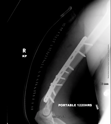
AP view after surgery and plating using 4.5mm plate and 3 lag screws


Lateral view after the placement of the plate

5 months after surgery the fracture is completely healed as shown below.


Often times it seems that the appropriate fixation is stabilization of the fracture with eight cortices of fixation and a minimum of three to four screws inserted proximal and distal to the fracture. 
It seems more accurate to take into consideration the concept of the working length of the plate. According to this concept increased fixation strength is noted with placement of screws close to the fracture, followed by plate fixation with screws spaced over a longer length plate. The working length of the plate may be more important than the number of cortices of fixation, and the increased spacing between screws provides advantages.

In the case described above three screws were spaced out proximally while fixation of the distal fragment was achieved with two plate screws and 3 inter-fragment lag screws.


A similar case which is more challenging is depicted below. The large butterfly fragment makes this fracture highly unstable and difficult to control rotationally. Placement of a mini plate or lag screws and conservation of the three part to a two part fracture may be necessary for control of the reduction and biomechanical stability of the fracture. The challenge with the segmental fractures at this level of the humerus is that the lag screws are difficult to be placed from lateral to medial direction as they may endanger the ulnar nerve. Mobilization of the ulnar nerve may be necessary. In addition, in muscular individuals, as in this case, the triceps splitting approach poses an obstacle to the placement of the lag screws as the bulk of the retracted medially or laterally triceps muscle (splitting approach) blocks or forces the screwdrivers or drills posteriorly. This "technical" limitation forces the trajectory of the lag screw in a rather oblique than parallel to the coronal plane direction on the bone. This is not ideal when the distal humerus is split in half on the sagittal plane as in this case.  Remember that the humerus is flattened distally which makes lag screw placement more difficult as described above.


Further reading:
Management of Humeral Shaft Fractures
J Am Acad Orthop Surg July 2012 ; 20:423-433