Monday, August 25, 2014

Glenoid vault penetration in anatomic total shoulder arthroplasty

There is often a lot of discussion and criticism about the position of the glenoid component in total shoulder arthroplasty. It is a critical step in total shoulder replacement surgery because the main reason for failure of a total shoulder arthroplasty is glenoid component loosening/failure. Without a doubt the placement of a glenoid component requires experience and consideration of many factors especially in the deformed arthritic osteopenic glenoids. Factors to take into consideration during reaming and placement of the glenoid component are:

(1) Degree  of the glenoid deformity. Correction or no correction of the deformity?
*(2) Reaming to subchondral or pass the subchondral plate? It seems that the implanted glenoid component is mostly supported by the subchondral bone
(3) Sizing of the implant. Larger or smaller implant?
(4) Use of step cut glenoid component or not
(5) Penetration of the glenoid vault or not?

In the case example provided below a preoperative CT scan demonstrates no glenoid deformity and implantation of a anchored pegged glenoid component was performed without penetration of the glenoid vault. It seems that pressurization of the cement in intact glenoid vault would be ideal if there is no cortex penetration. Although it is critical when keeled glenoid components are used it seems that it has no implications when anchored pegged glenoid components are used. The reason is that we do not use cement for the central peg. In case of penetration it seems that for the central anchored ingrowth peg there will be "bicortical fixation".

Further reading:
Edge displacement and deformation of glenoid components in response to eccentric loading. The effect of preparation of the glenoid bone. D Collins, A Tencer, J Sidles and F Matsen. J Bone Joint Surg Am. 1992;74:501-507

Central anchored peg contained in the glenoid vault


Grashey view shows the height of the prosthesis and alignment

Preoperative CT scan shows no glenoid deformity.


The ultrasonographic appearance of biceps tendinitis

The argument about the use of ultrasound in musculoskeletal imaging is that is operator dependent. Studies show that the accuracy in diagnosis is affected by the experience of the sonographer. Although it has not replaced the use of MRI in the evaluation of the rotator cuff and labrum,  without a doubt the use of sonography is gaining ground in the evaluation of shoulder. We routinely use MSK ultrasound for the evaluation of shoulder problems because 1. It is a dynamic examination of the soft tissues 2. It is cheap 3. It is fast and can be done in the office 4. It improves the accuracy of cortizone injections.
The images below demonstrate fluid around the biceps tendon in the bicipital groove indicative of biceps tendinitis


Probe position
Prior to injection the biceps in the bicipital groove is surrounded by fluid (tendinitis)
Using sonographic guidance cortizone injection was given to the biceps tendon sheath showing the expansion of it


Saturday, August 9, 2014

Technical tips for operative treatment of segmental both bone forearm fractures

The following case was a high energy trauma victim that was taken to the operating from the emergency department. This is complex fracture involving the forearm. There was a segmental radius fracture with an associated distal ulnar shaft fracture. The technical tips for this surgery are the following:
-The radius was exposed through an extensile volar Henry approach. Start the dissection distally, keeping the FCR and the radial artery retracted medially and extend the interval proximally as needed. Distally it is easier to identify the structures. Keep in mind that in the classic Henry approach the FCR and radial artery are retracted medially. This ensures that the radial artery will be on the medial side when the incision is extended proximally. In the modified Henry approach the FCR is retracted medially and the radial artery laterally. If a modified approach is used then when you extend the incision proximally the radial artery will be crossing the surgical field as you develop the brachialis and FCR interval
-Pay attention to the bow of the radius and make every effort to restore it.
-Convert the three segments to two segments with a use of a lag screw when possible. That facilitates anatomic alignment of the fracture
-Most plates that are not precontoured will need bending to fit the radius.
-Use 3.5mm non locking plates for no articular fractures
-Fixation of the intermediate segment requires two screws for rotational control when one plate is used for fixation of the segmented bone. If two plates are used then you need two screws for each plate applied to the intermediate fragment
-The working length of the plates is more important for biomechanical stability than the total number of screws used
-Partial or complete release of the supinator off the radius proximally may be required. If a complete release is performed leave a cuff of tendon for repair at the end
-Keep the forearm in supination to avoid iatrogenic injury to the posterior interosseous nerve proximally.

Xrays prior and after the fixation of the fracture are shown below.


Friday, August 8, 2014

Distal humerus fractures in the elderly

There is an increasing concern about the distal humerus fractures in the elderly. The concern is about the increasing frequency of this injury in patients with poor bone stock. These patients are usually in their 80s or 90s and pose difficulties in their treatment due to the high levels of activity combined with challenging injuries around the elbow. Open reduction and internal fixation remains the treatment of choice. However, when there is poor bone density fixation can be challenging and the option of total elbow replacement should be entertained. In the years to come orthopaedic surgeons will be doing a lot more total elbow replacements for the reasons explained above. 

The following case is an elderly patient who presented with a distal humerus fracture after a fall. She is physiologically a lot younger than her chronologic age and decided to proceed with operative treatment of the fracture. We prefer, when possible, to do "a triceps on" surgical approach and avoid olecranon osteotomy. The trans-olecranon approach is mandatory when there are multiple intra-articular fragments that require visualization for anatomic reduction and restoration of the smoothness of the articular surface. Xrays before and after surgery are shown and the options of parallel versus biplanar plating are discussed below. In this case two lag screws and orthogonal plating was used for the 3 part distal humerus fracture


Parallel plating
There may be times when it is not possible, because of the specific anatomy of a fracture complex, to place the lateral plate posteriorly, and it has to lie on the crest of the lateral column. In such a scenario, the planes of the two plates will be parallel.
Some surgeons use parallel plating as the preferred method, on the basis that a posterolateral plate permits only short unicortical screws distally, whereas a lateral plate allows the use of longer distal screws.
The choice of method will be determined by the bone quality, the fracture anatomy, the availability of locking plates and individual surgical philosophy.

Biplanar plating
The use of two plates on the distal humerus for C-fractures greatly assists the reconstruction of the triangle of stability. 
When one plate lies on the crest of the medial column, and the other lies on the posterior aspect of the lateral column, which is nonarticular, the two plates lie in planes that are at 90 degrees to each other – perpendicular, or biplanar, plating. 
Such a construct is considered to confer good biomechanical stability in good quality bone, but this has not been proven experimentally.

Wednesday, July 2, 2014

The different faces of shoulder arthritis. Intra-operative pictures

The following case is an elderly woman with many years of pain and crepitus to the shoulder. Her active forward elevation was to 50 degrees with intact rotator cuff. The patient failed conservative treatment and elected to have a total shoulder replacement for her shoulder arthritis. Preoperative, intra-operative and postoperative images are shown below. Most of the time we find loss of articular cartilage and flattening of the humeral head. In this case the arthritic changes were cystic changes to the humeral head and glenoid. Of note the patient has no history of autoimmune disease or inflammatory arthropathy.

Preoperative on the right and postoperative image on the left

Left: Axillary view shows no dislocation. Right: Erosive changes to humeral head and glenoid are seen

Cystic changes to the humeral head were seen intra-operatively

No cartilage was found on the glenoid

Tuesday, July 1, 2014

When do we have to fix clavicle fractures?

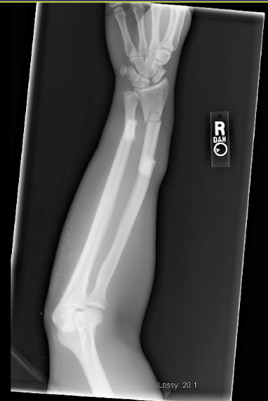
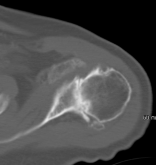
This case is a patient in his 30s who is construction worker (high demand) and was seen in the office due to his R clavicle fracture. He presented to the office with R clavicle pain due to a fracture that was 5 weeks old. He was concerned about his ability to return to work after healing of this fracture in conservative fashion ie treatment in a sling. Xrays demonstrated a displaced clavicle fracture with more than 2 cm of shortening. We elected to proceed with open reduction and internal fixation due to his profession and the radiographic appearance of the fracture. Xrays prior and after the fixation as shown below. In addition, the evidence for surgical approach for this type of injury is reported below (AAOS)


Do all displaced midshaft clavicle fractures need surgery?

By Jennie McKee
Point/counterpoint: ORIF or nonsurgical treatment
Whether an athlete falls off a bicycle, is tackled in a football game, or is slammed into the side of the hockey rink by another player, the result can be the same: a displaced midshaft clavicle fracture. When this potentially season-ending injury occurs, it’s not always clear whether conservative treatment or surgical fixation will yield the best patient outcomes.
Brian H. Mullis, MD, and Edward G. McFarland, MD, explored the pros and cons of both treatment strategies during a point/counterpoint session at the 2011 Specialty Day meeting of the American Orthopaedic Society of Sports Medicine.
Benefits of surgical fixation
According to Dr. Mullis, treating displaced midshaft clavicle fractures with open reduction and internal fixation (ORIF) can offer several benefits compared to conservative treatment, because athletes can get back on the field sooner.
In a prospective, randomized, multicenter study of more than 100 patients with displaced midshaft clavicle fractures, for example, the nonsurgical group healed at an average of 28 weeks, while the surgical group healed at an average of 16 weeks.
“I’m assuming most patients would like the much shorter healing time that surgical fixation provided in that study,” he said, adding that surgery offered other benefits as well.
“Researchers found that patients treated nonsurgically had higher malunion and nonunion rates and worse outcomes,” he said.
Those conclusions were reinforced by another prospective, observational study that also found higher nonunion rates in patients treated nonsurgically.
“The study found a 30 percent nonunion rate and a 36 percent symptomatic malunion rate in the nonsurgical group,” he said, “while the surgical group did much better.” He added that both the Constant and the American Shoulder and Elbow Surgeons (ASES) scores were worse in the nonsurgical group at all time points in the study.
Strength in the affected limb can also be an issue with nonsurgical treatment, he said. Although many patients likely have good long-term results when treated nonsurgically, some studies have found a correlation between nonsurgical treatment and decreased strength.
He noted that a 5-year follow-up study found that patients had a 20 percent to 30 percent loss of strength on the injured side compared to the uninjured side. They also had much worse functional scores on the injured side.
Indications for surgery
“Absolute indications for surgery include open fractures and neurovascular injury requiring repair or exploration,” said Dr. Mullis, who suggested that the strongest relative indication for surgery is a displaced clavicle with 2 cm or more of shortening (Fig. 1). Other relative indications include multiple extremity involvement, floating shoulder, seizure disorders, and cosmesis.
SSM_Pt-Ctrpt_Fig 1A.gif
SSM_Pt-Ctrpt_Fig 1B.gif
Fig. 1 (From top): Pre- and postoperative radiographs of a patient who underwent surgical fixation of a displaced midshaft clavicle fracture. Courtesy of Brian H. Mullis, MD
“I’ll be the first to admit that for an athlete with a truly nondisplaced fracture, the risks of surgery outweigh the benefits,” said Dr. Mullis, “and I certainly would not provide surgical treatment for children, who heal well with nonsurgical treatment. I would also not recommend surgery for patients with low functional demands.”
ORIF is “not without its perils”
Like Dr. Mullis, Dr. McFarland believes that ORIF can be the best treatment option for displaced midshaft clavicle fractures in competitive athletes early in the season, but cautions that not every clavicle fracture needs surgical fixation.
“The question a treating orthopaedist should ask is not, ‘Should I surgically repair this injury in the athlete?’ but ‘Is this the type of fracture I would repair surgically in any patient?’”
Variables such as degree of displacement, shortening, and comminution should be carefully considered. Other variables include location of the injury (dominant or nondominant arm), the patient’s activity level, status of the physes (open or closed), and the existence of other injuries.
“An orthopaedist needs to reconsider the advantages of giving that patient 2 or 3 extra weeks to compete, if, for example, the patient is not making a living from a sport or not involved in sports at all,” he said.
He agreed with Dr. Mullis that children with open physes generally should not undergo surgical fixation.
Surgical complications
“Whenever you do operate on patients with this injury, you have to weigh the benefits and the risks,” said Dr. McFarland. “Incisional numbness and hardware irritation are real and not infrequent complications, as are scar concerns and the risk of infection.”
Studies support the risk of surgical complications. In one study, 53 percent of surgical patients required plate removal. In another study of 125 patients who underwent ORIF, 12 percent needed reoperation, 4 percent had plate breakage, and 3.2 percent had loosening. Other complications included infection and frozen shoulders.
“This tells us that ORIF is not without its perils,” he said. “And the question is, does it really give us that much of an advantage?”
Studies have shown, said Dr. McFarland, that healing time with a clavicle reconstruction plate is 14.6 weeks; healing time with a compression plate is 13 weeks. A study of professional football players found that nonsurgical treatment yielded a healing time of 7 weeks, compared to a healing time of 8.8 weeks with ORIF.
“And, remember, the sooner the patient returns to sport, the greater the risk that another injury might occur,” he added.
Some studies suggest, said Dr. McFarland, that providing conservative treatment and waiting to see if a nonunion occurs, rather than initially treating the patient surgically, may not pose significant risks. He pointed to a study that found that if nonunions are operated on later—meaning, after the patient has undergone conservative treatment and subsequently required surgical fixation for a nonunion—the results are almost the same as with immediate ORIF.
“I tell most of my patients—even those with displaced fractures—not to be concerned about a nonunion, because if it does occur, we will repair it surgically later, and they will be as good as they were before,” he said.
In closing, Dr. McFarland emphasized that treatment should be individualized to the patient and the fracture pattern.
“ORIF in midseason is a viable option in some athletes,” he said, noting that patients must be educated about the possibility of complications and the need for reoperation. Despite these risks, he said, “surgical fixation can be satisfying in the right patient.”
Dr. McFarland acknowledged the assistance of Juan Garzon-Muvdi, MD, in preparing his presentation.
Disclosure information: Dr. Mullis—Synthes, Amgen, AO, Medtronic. Dr. McFarland—Stryker DePuy-Mitke; DePuy, A Johnson & Johnson Company; Stryker; and DJ Orthopaedics; American Journal of Sports Medicine; Journal of Athletic Training; Clinical Orthopaedic and Related Research; Medicine and Science in Exercise and Health.
Jennie McKee is a staff writer for AAOS Now. She can be reached atmckee@aaos.org
Bottom Line
  • When treating displaced midshaft clavicle fractures, orthopaedists must take many variables into account, including (but not limited to) the degree of displacement, shortening, comminution, patient age (open epiphysis), and the existence of other injuries.
  • According to some studies, ORIF enables athletes with displaced midshaft clavicle fractures to return to sport sooner than conservative treatment.
  • Drawbacks of surgical fixation can include hardware irritation and removal, incisional numbness, infection, and scar concerns.
  • Although ORIF can be a valid option in appropriate patients, orthopaedists should ask whether this is the type of fracture that needs an operation in any patient before proceeding.
References:
  1. Canadian Orthopaedic Trauma Society: Nonoperative treatment compared with plate fixation of displaced midshaft clavicular fractures. A multicenter, randomized clinical trial. J Bone Joint Surg Am 2007;89(1):1-10.
  2. Kulshrestha V, Roy T, Audige L: Operative versus nonoperativemanagement of displaced midshaft clavicle fractures: A prospective cohort study. J Orthop Trauma 2011;25(1):31-38.
  3. McKee MD, Pedersen EM, Jones C, et al: Deficits following nonoperative treatment of displaced midshaft clavicular fractures. J Bone Joint Surg Am 2006;88(1):35-40.
  4. Smekal V, Irenberger A, Struve P, Wambacher MKrappinger D,Kralinger FS: Elastic stable intramedullary nailing versus nonoperative treatment of displaced midshaft clavicular fractures: A randomized, controlled, clinical trial. J Orthop Trauma2009;23(2):106-112.
  5. Ferran NA, Hodgson P, Vannet N, Williams R, Evans RO: Locked intramedullary fixation vs plating for displaced and shortened mid-shaft clavicle fractures: A randomized clinical trial. J Shoulder Elbow Surg. 2010;19(6):783-789.
  6. Potter JM, Jones C, Wild LM, Schemitsch EHMcKee MDDoes delay matter? The restoration of objectively measured shoulder strength and patient-oriented outcome after immediate fixation versus delayed reconstruction of displaced midshaft fractures of the clavicle.J Shoulder Elbow Surg 2007;16(5):514-518. Epub Jul 12, 2007.

Saturday, June 28, 2014

Reimplassage combined with Bankart repair for recurrent traumatic anterior shoulder instability.


Hill Sachs lesion at the posterior superior aspect of the shoulder

Placement of anchor and reimplassage


In French reimplassage means "to fill in". During this procedure the engaging Hill Sachs lesion of the humeral defect is filled in the infraspinatus tendon and the capsule. The Hill Sachs lesion is converted to an extra-articular lesion that does not engage with the anterior glenoid rim providing stability to the shoulder. The procedure was described in 2007 by Wolf et al as an adjunct to the arthroscopic anterior stabilisation procedure of the shoulder in order to address a large engaging Hill-Sach's defect.  

It is ideally suited to instability patients who have large, engaging Hill-Sachs lesions and soft-tissue Bankart tears. These patients are known to have a higher failure rate after surgery than those with smaller lesions. The results of this technique in this difficult subset of traumatic anterior shoulder instability patients are significantly better (10% recurrence rate) than with an arthroscopic Bankart repair alone (67% recurrence rate).

The following case is a patient is his 20s who had 30 dislocations during the past 6 years. His first traumatic anterior shoulder dislocation was a result of a wrestling accident. Due to his young age and no bone loss at the glenoid side we elected to proceed with an arthroscopic Bankart repair combined with a reimplassage procedure due to the large engaging Hill Sachs lesion. Imaging studies and arthroscopy pictures are shown below.

Grashey view

Apical oblique shows no glenoid bone loss

Bankart lesion; view from anterior and posterior portal

Repaired Bankart