Wednesday, August 16, 2017

The ideal position of the glenosphere in primary reverse total shoulder arthroplasty

The reverse shoulder arthroplasty is a highly technical operation. It requires appropriate soft tissue balancing and correct component positioning. When done for primary rotator cuff arthropathy has a very high success rate in restoring mobility and providing pain relief. When Paul Grammont introduced with procedure in France in the 1980s, the rest of the world saw it with scepticism and some orthopaedic surgeons had concerns. Today, it provides solutions for problems that we did not have a solution before. Below are the radiographs of a patient that has primary rotator cuff arthropathy, the patient is an elderly individual 70 years or older.


Postoperative radiographs show a press fit stem, inferior placement of the glenosphere and overhanging inferiorly by 2mm to avoid notching. Inferior tilting of the glenoid component is seen as well.
The superior screw at the 12 o'clock position is aiming towards the base of the coracoid, the inferior 6 o'clock screw is aiming to the spine of the scapula. The posterior screw is aimed inferiorly and anteriorly and the anterior screw superiorly and posteriorly. These are important details that achieve appropriate implantation and seating of the glenosphere which is the most critical aspect of this surgery.

The overhanging of the glenosphere by 2 mm avoids impingement of the polyethylene to the scapula

Correct position of the glenosphere


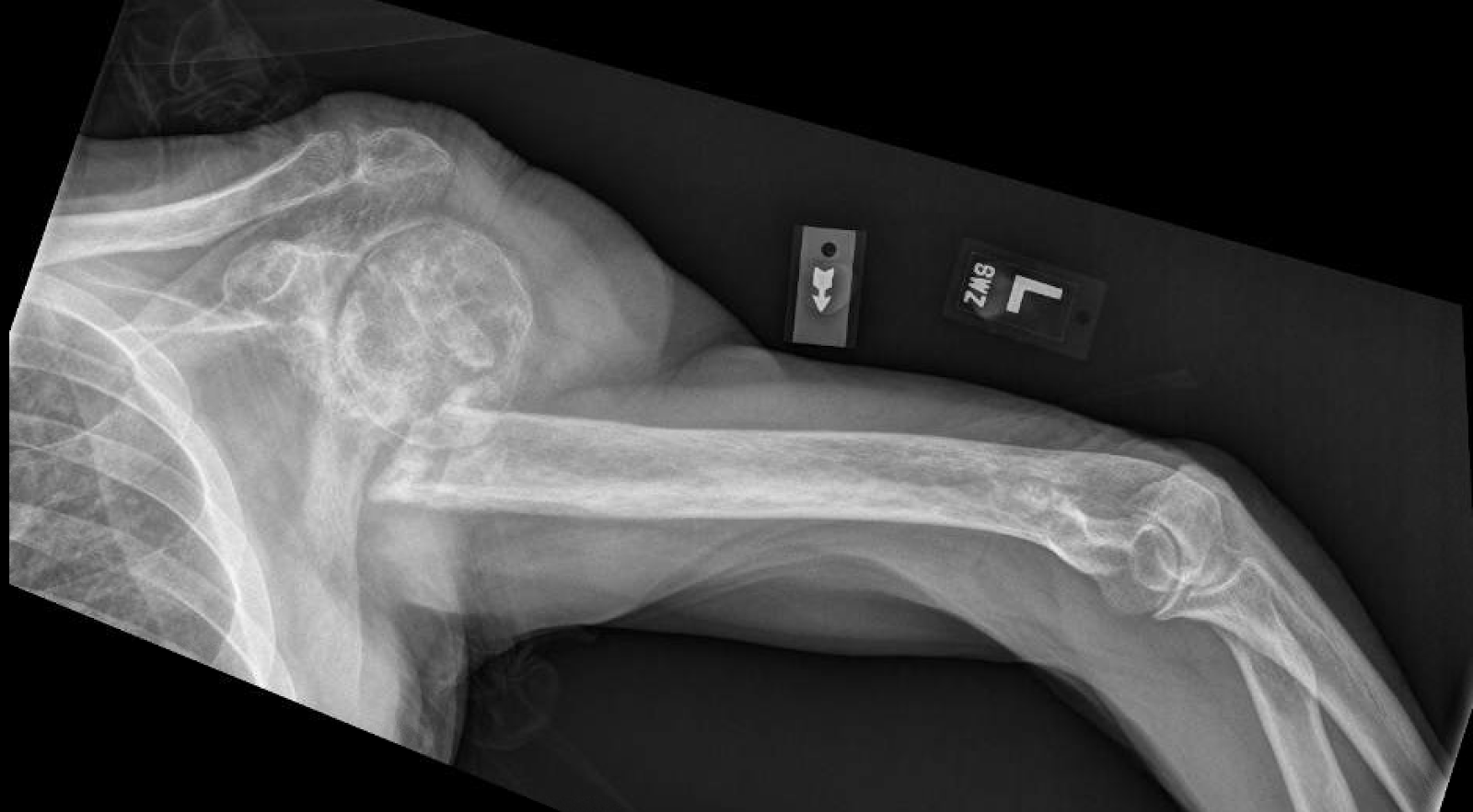
The axillary view shows  the appropriate orientation of the glenosphere.

Sunday, August 13, 2017

Eccentric prosthetic humeral head for Walch B2 arthritic glenoids.

The following case illustrates the benefit of using anteriorly directed eccentric head in anatomic arthroplasty to address the posterior subluxation of the humeral head seen often with a B2 arthritic glenoid. The details of the technique can be found by clicking here.


The deforming forces in proximal humerus fractures

Proximal humerus fracture fixation is challenging when there is insufficient bone density, more than 2 parts, an associated dislocation, and more importantly bone loss or impaction of the head in the setting of an obese patient or large deltoid which makes the exposure difficult. 

The deforming forces can be eliminated to a certain extend when the patient is placed in the supine position with the arm on a hand table and abducted to 90 degrees which relaxes the deltoid to allow for space retraction and application of the plate. The most significant deforming forces are:

1. Pectoralis major pulls the shaft anterior and medial
2. The greater tuberosity is pulled superiorly and posteriorly by the supraspinatus, infraspinatus and teres minor

Those forces can be significant and suture passing around the fragments can help with manipulation of the bone fragments. In the majority of cases the difficulty is in raising the articular surface, filling the defects of the bone without overstuffing and repairing the tuberosities. The head shaft angle is better tolerated in the valgus than in the varus position.

It is important to achieve the following after ORIF if possible:
a. Avoid long head screws as the most common complication is head screw penetration
b. Choose isolated screw fixation only in the young patient with good bone stock
c. Restore medial disrupted bone hinge
d. avoid step off in head splits
e. try to proceed with ORIF of head splits in young patients and avoid arthroplasty.
f. avoid superior migration of the greater tuberosity - or posterior superior migration - of more than 5 mm.
g. Pass sutures around the rotator cuff and tight them to the plate.
h. Avoid superior placement of the plate as it may impinge to the acromion with elevation of the shoulder or arm

Below is a fracture treated with ORIF taking into consideration those principles.


Proximal humerus fracture dislocation - Do we need to address the instability acutely?

This is a middle aged patient with a proximal humerus fracture dislocation. On the axillary view it is demonstrated that the humeral head is locked in a anteriorly dislocated position. The anterior inferior rim of the glenoid created the Hill Sachs lesion. The impaction was significant as the greater tuberosity is separated from the head and displaced and half of the glenoid is impacted in the humerus. Those injuries do have sometimes an associated soft tissue or bony Bankart lesion. Most of the time it is not necessary to address the instability at the time of fixation of the fracture, there is no need for repair of the soft tissues and no need for ORIF of the small glenoid fracture when it is present.

The stiffness that develops due to the proximal humerus fracture limits the ROM of the shoulder and for that reason the shoulder remains stable. Close follow up after surgery is recommended as such fractures can become unstable in the acute postoperative period.


Thursday, June 29, 2017

Arthroscopy journal: Technique for arthroscopic fixation of glenoid fractures

Abstract

The most common procedure to address transverse glenoid fractures that are characterized by intra-articular step-off or gapping is open reduction and internal fixation. Disadvantages of open surgery are delay in regaining full range of motion, increased approach morbidity, neurovascular complications, and the need for capsulotomy, which delays healing and increases the risk of stiffness. An arthroscopically assisted fracture fixation, as described in this article, is characterized by better visualization of the glenoid articular surface and reduction of the intra-articular fragments under direct vision, which diminishes the chances of residual step-off after fixation. Furthermore, arthroscopic fixation provides the advantages of minimal surgical trauma, which speeds up the recovery time, decreased morbidity as there is less blood loss compared with the open technique, lower chance of neurologic injury as there is less dissection around the spinoglenoid or suprascapular notch, less trauma to the joint capsule, and lower chances of stiffness and capsulorrhaphy arthropathy.

Anastasios Papadonikolakis, MD, PhD, Arthroscopy Techniques June 2017

Wednesday, June 28, 2017

Reverse shoulder replacement for chronic non union of the proximal humerus

The use of the reverse shoulder arthroplasty in the elderly is valuable not only for the treatment of rotator cuff arthropathy but also for the treatment of painful non union of the proximal humerus. My experience has been that open reduction and internal fixation is not a predictable solution in these cases due to the osteoporosis, poor nutrition and blow flow to the bone. This case below illustrates that the reverse total shoulder replacement provides solutions for problems that we did not have a solution before.

I prefer to use large glenospheres for those cases as instability can be a problem

I also prefer to use eccentric glenospheres which minimize the risk of impingement and scapular notching especially when the neck shaft angle of the humeral stem is more than 135 degrees. Those stems bring the humerus closer to the glenoid and can impinge. Impingement can be avoided by implanting a glenosphere with a 2-3 mm of inferior eccentricity.

The preoperative and postoperative images are shown below. This patient is in his 80s.

Dissection can be challenging especially when the proximal humerus is migrated in the axilla where the axillary a. and nerve can be traumatized at the time of surgery. Caution and patience is recommended during the dissection.


Friday, June 16, 2017

Arthroscopic findings after a ream and run shoulder hemiarthroplasty

Although the ream and run procedure has been a matter of debate among shoulder and elbow surgeons, it seems that for the young individual with glenohumeral arthritis there are not many alternative options. Glenoid components fail 10-15 years after surgery and some of these require difficult revision surgery as there is no bone left at the glenoid for reconstruction. The principle of ream and run surgery as developed by Dr Frederick Matsen at the University of Washington is that by reaming the glenoid to a smooth surface there is fibrocartilage regeneration, redistribution of the load and contact pressures to the entire glenoid and decrease in pain.

As reported by Dr Matsen:

"Earlier studies on hip and knee arthroplasty have provided encouraging evidence that reamed bone articulating with a convex metal prosthesis can remodel to a functional and durable arthroplasty concavity, sometimes lasting over four decades. Notably, the majority of so-called mold arthroplasty failures of the hip were on the femoral side and not on the acetabular side of the articulation. An analysis of pelvic specimens retrieved post mortem revealed that the concave acetabular joint surface was often covered with a smooth regenerated surface and had reestablished homogeneous and stable subsurface bone. Moreover, the tissue covering the acetabular concavity was found to resemble dermis and meniscus in terms of glycosaminoglycan content."


The case illustrated below is a of patient 1 year after a ream and run. During arthroscopy we found patchy formation of fibrocartilage on the glenoid side which probably accounts for the pain relief seen at the ream and run surgery. Pictures and video are demonstrated below.