Thursday, April 2, 2020

My online course - OrthoMesh3D: The use of CGI technology to plan corrective osteotomies and total shoulder replacement surgery


Description
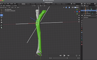
OrthoMesh3D


This course covers the basics of the free open source software named Blender, and how to utilize it in the planning of orthopaedic surgery using 3D bone models from CT scans. The modelling and animation of bones and joints will be covered, as well as the principles of deformity correction using cutting guides in the 3D environment using the Add on script - OrthoMesh3D. The OrthoMesh3D script is given to the students for free once they buy the course. Explanations are given on converting DICOM CT scans to 3D bone models, cleaning of bone models in Autodesk Meshmixer (free software) and importing them into Blender for surgical planning. There is a detailed explanation on how to use the free software Inkscape for converting pictures of implants to SVG files which then can be converted into 3D models in Blender. Lastly, a case example of a reverse shoulder replacement will be covered in addition to animation and simulation methods using the Blender's Armature tool. Inverse and Forward Kinematics for joint and bone model animation is covered too.
Who this course is for:
  • Surgeons, Medical Personnel, Engineers, Implant companies, Physicians, Medical Students
  • Python developers, Radiology physicians, Students or Instructors of Anatomy of the Musculoskeletal System

Wednesday, December 18, 2019

The posterior elbow Monteggia lesion, with associated instability of the elbow

Transolecranon elbow fracture dislocations, or posterior Monteggia elbow lesions are difficult injuries to treat. The following case illustrates the complexity in the fixation of a fracture that was 4 weeks old. The dual plating is necessary to reconstruct the ulna in those occasions. In addition, the fixation of the anteromedial facet of the coronoid is crucial to the stability of the elbow. In this case it was fixed with lag screws through the plate. About 2 years after surgery the patient had near complete ROM in all planes, however removal of the olecranon plate was necessary as the plate was painful.


Tuesday, October 29, 2019

Revision of failed anatomic total shoulder arthroplasty to reverse for subscapularis failure

In chronic ruptures of the subscapularis after anatomic total shoulder arthroplasty the dilemma arises. Should someone repair the subscapularis with a graft or convert to a reverse total shoulder replacement? If the patient is older than 65-70 years and is a low demand patient then the best option is a conversion to a reverse. The following example illustrates such approach. The patient regained full ROM after the revision. Prior to the revision her active forward elevation was to 50 degrees.


Wednesday, July 17, 2019

Open reduction and internal fixation of a 4 part proximal humerus fracture


ORIF of the proximal humerus can be challenging due to the several deforming forces applied by the muscles to the bone fragments. In addition, the deltopectoral approach provides a good exposure of the anterior, medial and anterolateral aspect of the proximal humerus but the posterior and posterolateral aspect can not to assessed adequately especially if the patient is muscular or obese.

It is important to have anatomic reduction and stable fixation of all the fracture fragments. It gives the patient the best chance of restoration of function to the shoulder. The illustrated case below is an example of a highly displaced and multi fragmentary proximal humerus fracture that was fixed with a locking plate and intramedullary fibular allograft. During surgery one of the fragments as indicated by the CT scan below was found be have been"driven" during the injury into the cancellous bone of the humeral head indicating high energy trauma.

Although an anatomic reduction of the fracture was achieved and eventual healing without complication the patient had active forward elevation only to 90 degrees at 12 months after surgery. There is no pain.

This case indicates that other factors may play a role in the functional outcome of this injury that are not related to "how well the fracture is reduced".


Friday, January 11, 2019

Does a stemless shoulder arthroplasty offer any benefits?

The stemless shoulder arthroplasty also known as resurfacing has gained popularity. When a new implant is introduced two basic questions need to be answered to identify its value:

a. What problem does the new implant solve? or "why is it superior to the existing implants?"
b. How "big" is that problem?

The theory in the development of stemless prostheses for shoulder replacement is the advantage of "bone preservation" and the ability for easier future revision. What we know today is that most shoulder arthroplasty constructs fail at the glenoid (glenoid component loosening) and that the humeral stem is rarely the reason for revision. Thus, to answer the second question of how big the problem is, it is probably insignificant.

It terms of the first question it seems that a smooth standard stem which is press fitted or a small size stem prosthesis with porous coating is rarely a problem during revision shoulder arthroplasty when extraction is required. For that reason, the theory of bone preservation is not very convincing. The indication, in my opinion, of using a stemless implant is when there is an existing metaphyseal proximal humerus malunion and implantation of a stem is problematic due to deformity of the humeral canal.

The problems that we see with the stemless shoulder implants are the following:

a. when the shoulder is painful it is nearly impossible to identify radiographically if the stemless implant is loose

b. In the Scandinavian and Australian registries they have higher revision rates compared to standard length hemiarthoplasties

c. Most of these implants are malpositioned due to the difficulty in establishing the neck shaft angle. An intramedullary "rod" is not used and frequently there is varus positioning as in the case below.

This patient had pain for 5 years after a Copeland resurfacing hemiarthroplasty. Radiographic analysis as shown below illustrates two problems:

a. the neck shaft angle indicates 124 degrees of inclination when the normal is 130-140 degrees. When the average neck-shaft angle of the 2058 humeri was calculated, 77% of the humeri had an angle of between 130 degrees and 140 degrees. Although,  the effect of a varus or valgus alignment of the prosthetic humeral head on the center of rotation of the articular surface seems small, it is unknown what the effect in the long term is.

b. There is 'overstuffing' of the shoulder joint which increases contact forces within the joint and tension to the rotator cuff leading to stiffness and pain which was the main reason that this patient was referred to us.


Neck shaft angle of 124 degrees


Center of rotation is medialized indicating overstuffing

axillary view indicates medialization of the humeral center of rotation


Revision to anatomic total shoulder arthroplasty with neck shaft angle of 134 degrees

Restoration of the center of rotation of the humerus


After the revision surgery to a total shoulder arthroplasty the patient had 160 degrees forward elevation, no pain and was pleased with the outcome at 6 months postoperatively.

Tuesday, January 8, 2019

Hemiarthroplasty for proximal humerus fracture

It is currently widely accepted that a four part proximal humerus fracture in the elderly is more reliably approached with a reverse total shoulder replacement (rTSA) compared to a hemiarthroplasty. There is argument that the rTSA provides better relief of pain and that if the tuberosities do not heal after a hemiarthroplasty then the outcome is poor in terms of motion of the shoulder. 

In the case illustrated below a hemiarthroplasty was chosen as the patient had balance problems and potential falls can be more forgiving for the hemiarthroplasty when compared to a reverse. A reverse shoulder replacement has a dislocation rate of up to 40% when done for fracture.

The Grashey xray shows consolidation of the bone graft in the "window" of the stem proximally and although someone may say that the greater tuberosity did not heal, it did heal but it is healed posterolaterally due to the pull of the infraspinatus. The proof is on the axillary x-ray shown below.

At 6 months the patient had no pain and active forward elevation to 140 degrees.

Individualizing the approach is the key to success for these cases although it is difficult to predict the outcome.


FDA Class 2 Device Recall - Smith and Nephew PROMOS Inclination set

This is a rare complication of anatomic shoulder arthroplasty. Dissociation of the humeral component from the stem is very rarely seen, especially when the fixation method is a morse taper. The following case illustrates the problem of "modularity". The more complexity is added to the prosthesis the higher the chances of failure of the implant. This patient had a Smith and Nephew Promos prosthesis implanted in his shoulder and he did well for quite some time, until he felt a snap at approximately 10 years after surgery. The following x-rays were taken in our office at the time of first evaluation which indicate a dissociation of the stem from the head. The FDA has issued a Class 2 Device Recall for this particular implant.


As seen in the picture above this design can have multiple modes of failure at the head / stem interface due to the complexity of the fixation. In addition, the entire stem is porous coated which makes it extremely difficult to revise and unnecessary as non coating or proximal coating is enough for fixation of the humeral stem and press fitting.

The idea in this case was to avoid conversion to a reverse TSA as the glenoid component was well fixed and there was intact rotator cuff at the time of surgery. Such a revision would have been very difficult as removal of the glenoid fixed component can lead to glenoid bone defect and extraction of the stem would have required extensile humeral osteotomy as it was porous coates in its entire length


The picture above shows the broken screw at the inclination set


The picture above shows the improved design/inclination set that was used for salvage of the construct.
Luckily, the retained broken screw was removed from the stem without damaging the threads of the stem

The patient had the least aggressive approach, which was to downsize the humeral head, retain the glenoid component, as it was well fixed despite the radiolucent lines and implant the new small head with the new inclination set.


At 6 weeks postop the patient had 170 assistive forward elevation, no limitation in internal rotation and no pain.