Saturday, May 3, 2014

Audience for shoulderelbow.blogspot.com. Thank you!

I would like to thank the users from North America, Russia, Latin America, Europe (especially Greece, Germany and France) and Asia for visiting my blog. It is greatly appreciated. Below is a map with color intensity based on frequency.

Best regards,
Anastasios Papadonikolakis, MD, PhD

Management of anterior fracture-dislocations of the proximal humerus. Is Bankart repair necessary?

In cases of un-reconstructable proximal humerus fractures or 4 part proximal humerus fractures in the elderly prosthetic replacement is recommended. The fracture dislocations are often times associated with glenoid rim fracture which most of the time are less than 20% of the glenoid. Generally, biomechanical studies as well as clinical studies have shown that glenoid bone loss of more than 20% needs to be addressed either with fixation of the fractured glenoid or with bone augmentation of the glenoid. In the setting of a proximal humerus fracture dislocation it is uncommon to have more than 20% glenoid bone loss because most of the energy of trauma is absorbed by the fractured humerus. However, it is common to find during surgery small glenoid rim fractures. These fractures do not need to be addressed because they do not lead to instability. Fixation of the bony bankart lesion will lead to stiffness.

In younger patients (age <65) and with 2 or 3 part proximal humerus fracture with viable humeral heads every effort needs to be made for fixation of the fracture. The following case is a 62 year old nurse who presented after a fall with an anterior fracture dislocation of the shoulder. The humeral head was dislocated anteriorly and there was a small bony bankart lesion at the anterior inferior aspect of the humeral head. Disimpaction of the fractured humerus from the glenoid can be challenging and in this case was performed by a horizontal split of the subscapularis muscle belly, insertion of an elevator between the glenoid and the fracture humeral head to assist with the reduction maneuver. Open reduction and internal fixation was performed. At 2 years postoperatively the fracture is united, the patient had an active shoulder forward elevation of 120 degrees on the injured side and was pain free.

Further reading: http://www.bjj.boneandjoint.org.uk/content/88-B/4/502.full


Friday, May 2, 2014

Repairing rotator cuff tears at the time of anatomic total shoulder replacement

Repair of a rotator cuff tear at the time of anatomic total shoulder replacement is critical. Patients with osteoarthritis of the shoulder that have no symptoms of rotator cuff deficiency may have at the time of surgery rotator cuff tears that need repair. Prior to surgery the patients are examined for obvious symptomatic rotator cuff tears, however if the surgeon encounters a torn cuff at the time of anatomic shoulder replacement then it is mandatory to proceed with repair. Such a repair will slow down the recovery and rehab protocol of the patient at the benefit of decreased chance of early loosening and failure of the glenoid component. For further reading please refer to:


The following case is a 70 y/o female with a long history of shoulder pain due to glenohumeral osteoarthritis. She failed conservative treatment and at the time of presentation she had intact rotator cuff strength. Preoperative radiographs indicate degeneration of the footprint of the supraspinatus with formation of the a large osteophyte at the footprint of the supraspinatus. In addition the so called "bone spur" is visible on the acromial side. Although this radiographic finding is indicative of degenerative tear of the supraspinatus it needs to highlighted that it not diagnostic. At the time of surgery she did have a 2 cm tear of the supraspinatus that was repaired. We elected not to remove the osteophyte because this would have required violation of the intact fibers of the supraspinatus. She was pain free at 4 months postop. Preoperative and postoperative Xrays are illustrated below. It is important to educate patients that "bone spurs" are not always the cause of pain.


Technical errors in anatomic total shoulder replacement

The anatomic total shoulder replacement is an operation that requires accuracy and technical skills. The balancing of the shoulder and correct placement of the implant are essential for a good outcome. A few of the most common technical errors are outlined below
(1) Malpositioned stems: The most common error is varus placement of the stem or high "sitting" of the stem that lead to "overstuffing" of the joint and high tension to the rotator cuff respectively.
(2) Injury to the infraspinatus during the humeral cut
(3) Over reaming or eccentric reaming of the glenoid in an attempt to correct the glenoid deformity. Correction of the glenoid deformity has the significant disadvantage of removing subchondral bone which is essential for support of the glenoid. This may lead to early loosening and failure of the glenoid component compomising the longevity of the reconstruction.
(4) Failure to adequately utilize the advantage of the eccentric heads to reproduce the anatomy of the proximal humerus
(5) Inadequate inferior capsule release during exposure that leads to difficulties in accessing the glenoid for reaming
(6) Incomplete seating of the glenoid because of poor reaming technique
(7) Over-reaming of the humerus that removes endosteal bone leading to stress points around the tip of the stem and subsequent periprothetic fractures
(8) Less than 4 sutures for repair of the subscapularis tendon that is associated with high failure of the repair.

 When the humeral cut is aimed inferiorly then there is a chance of injury to the infraspinatus
 Superior prominence and medial displacement of the implant leads to "overstuffing" and poor outcomes

 Eccentric placement of the canal was difficult to correct in the past prior to the introduction of the eccentric heads. Minor deviations from the anatomic placement can be corrected with eccentric heads. We recommend identification of the insertion of the supraspinatus and placement of the stem just a few mm medial to the insertion to avoid varus deformity and eccentric placement

Thursday, May 1, 2014

Reverse Bankart lesions and shoulder instability

This is a 25 year old male who was seen in the office with a two year history of shoulder pain. He had tried physical therapy without resolution of this pain. He reported that 2 years ago he was bench pressing at the gym when he felt sharp pain in the shoulder. On exam he had a positive external rotation shear test and a positive Jerk test not for instability but for pain in the periscapular area. He denied instability or dislocations. Radiographic evaluation of the shoulder with plain films was unremarkable for pathology. At the time of referral he already had a CT scan and MRI of the shoulder.


The CT scan demonstrated what is reported in the literature as a Bennett lesion indicative of posterior instability of the shoulder or internal impingement of the shoulder. A Bennett lesion is a bony spur that usually forms at the posterior inferior glenoid rim and sometimes becomes painful. Most of the time detachment of the posterior glenoid labrum is seen during arthroscopy associated with posterior capsular tightness.

The CT scan shows the erosion and bony spur at the posterior rim of the glenoid

MRI indicates an associated posterior labrum tear.

The patient underwent a program of 6 weeks of posterior capsular stretching without improvement of his symptoms. He was taken to the operating room for arthroscopic repair of his labrum and removal of the unstable bone or cartilage fragment that was unstable and has been related in the literature with recurrence of the symptoms.

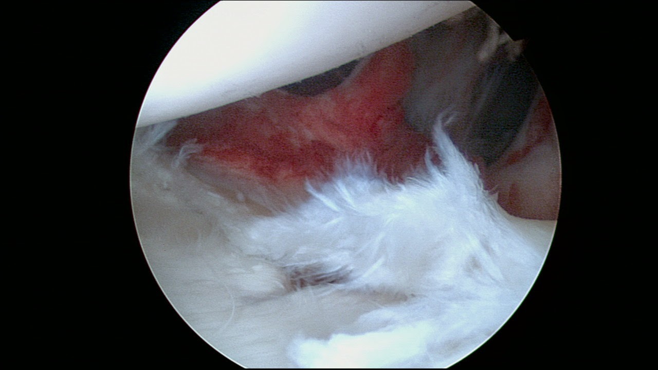
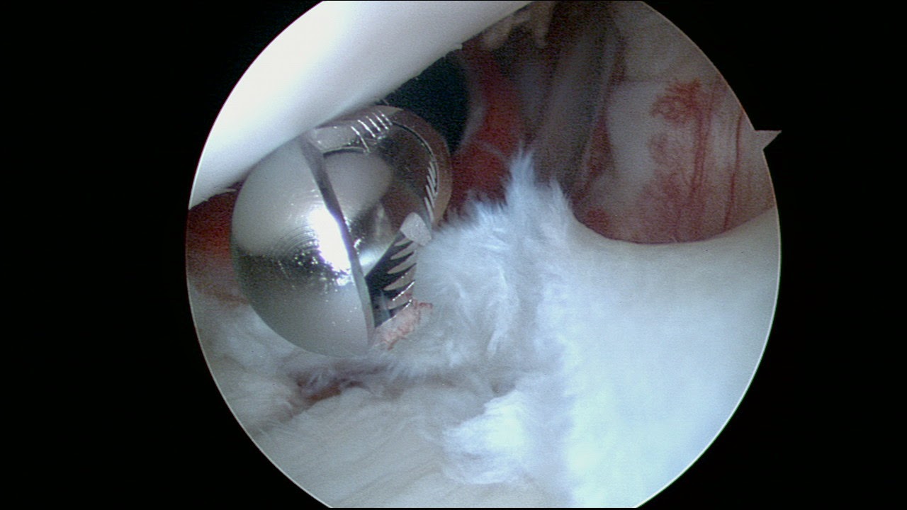
The following pictures demonstrate the cartilage defect after removal of the lesion and the labrum tear. Viewing from the anterior portal.


 Final repair of posterior labrum pathology
The patient followed a extensive program of physical therapy and returned to sports 6 months after this repair without pain.


Proximal humerus nonunion in the elderly - reverse shoulder replacement

The following case illustrates the use of a reverse shoulder replacement in the treatment of a long standing nonunion of the the proximal humerus. This patient has a 4 year hx of proximal humeral surgical neck fracture was treated elsewhere in a conservative fashion. She is a heavy smoker and otherwise indepedent ambulator and very active. She is 68 y/o F who had at the time of presentation active forward shoulder elevation to 30 degrees. She had severe pain and was treated by pain management. Her deltoid muscle was intact.


This radiograph demonstrates cavitation of the humeral head longstanding nonunion of the proximal humerus. 
The treatment options are (1) Osteosynthesis with bone grafting (2) Prosthetic replacement.

We elected to proceed with a reverse shoulder replacement due to the smoking status and the age of the patient. 


At 6 weeks postop she had minimal pain and assistive forward elevation to 140 degrees.

Open reduction and internal fixation with bone grafting would have been the treatment of choice for a patient of younger age and no smoking status.
A reverse total shoulder replacement has the advantage of earlier initiation of motion however it does have the disadvantages of glenoid failure which can be up to 30% at 10 years after surgery as well as the risks of complications related to prosthetic joint replacement.

The patient returned at 2.5 months with full active ROM to the R shoulder 


Distal clavicle fracture fixation using locking plates

During a previous post it was illustrated that non locking 3.5 reconstruction plates are sufficient for fixation of clavicle shaft fractures. However, distal clavicle fractures are challenging and they require multiple screws for the distal fragment. Re-enforcement of the fixation with suture that is wrapped around the coracoid as almost always necessary. These fractures are more challenging and require compliance with restrictions during the healing period to avoid biomechanical failure of the fixation.

The following case is a 30 year old M who presented to the office with a distal clavicle fracture after an altercation. Fixation was performed with a distal locking 3.5 plate and the reduction of the superior displacement of the clavicle was re-enforced with the use of #5 Fiberwire suture what was placed around the base of the coracoid. The pre-operative planning was essential for the use of a plate that allows placement of multiple distal locking screws.


3D reconstruction and templating 


intraop images


Immediate postop images

4 locking distal screws and suture fixation for the distal fragment

Preoperatively (L side is the injured side)

Postoperatively