Wednesday, September 17, 2014

Shoulder hemi-arthroplasty for humeral head avascular necrosis

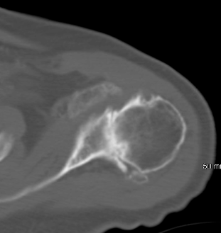
The following case is a middle aged patient with shoulder pain for several years. The radiographic evaluation of the shoulder demonstrated post-collapse avascular necrosis of the humeral head. The cause of his disease was alcohol abuse. While some surgeons believe that a "minimallly invasive" procedure is appropriate for the treatment of this condition we avoid the use of such procedures because:
(1) mini caps have the problem of loosening and do not prevent further progression of the disease

(2) shoulder resurfacing implants result in early loosening and revision to hemi-arthroplasties.

The longevity of a hemi-arthroplasty is approximately 10 years before glenoid wear becomes an issue. At that point a conversion of the hemi-arthroplasty to a total shoulder replacement can be offered extending the longevity to an additional 10-15 years.  Preop/postop radiographs and intraoperative images are shown below


Severe loss of cartilage from AVN


Tuesday, September 9, 2014

Type I-II-III-IV pediatric supracondylar humerus fractures

The majority of pediatric supracondylar humerus fractures are extension type injuries. Fixation of these fractures requires attention to detail and avoidance of use of medial pinning when possible. Use of medial pins runs the risk of injury to the ulnar nerve and there is no additional biomechanical stability by adding medial pins. Gartland classified these fractures in 3 types based on the displacement and treatment method.

Gartland Classificaiton 
Type INondisplaced, beware of subtle medial comminution leading to cubitus varus
Type IIDisplaced, posterior cortex intact
Type IIICompletely displaced
*Type IVComplete periosteal disruption with instability in flexion and extension

Type I fractures can be treated in a long arm cast or with pinning. Some orthopaedic surgeons prefer pinning of the non displaced  Type I fractures in an attempt to avoid lost to followup cases, avoid frequent visits to the office and radiation exposure. Currently, both methods of treatment are acceptable as long as a close weekly followup is scheduled for the patients that receive conservative Rx in a cast without pinning. If the fracture displaces then pinning is required. 

The case below is a 4 year old patient who presented with elevation of the fat pad on the lateral view due to a non displaced supracondylar humerus fracture. She was treated in a long arm cast for 6 weeks and healed without complications.


The following case is a 3 year old patient who presented with a Type II supracondylar humerus fracture that was treated with 2 lateral pins. The fracture healed without complications at 6 weeks.


The last case is a 4 year old patient who presented with a Type III supracondylar humerus fracture and was treated with lateral pinning only. The patient had the pins removed at 3 weeks and received long arm cast immobilization for a total of 6 weeks. Xrays show a healed fracture at 6 weeks.


XRs 6 months after show healed fracture. XRs are shown below at 6 months after surgery.


During fixation of these fractures it is ideal to have tricortical fixation by penetrating the coronoid fossa with the pins as demonstrated in the picture below.


This method of fixation provides better biomechanical and rotational control of the fracture. It is important to take into consideration the 3 column theory and private stability to the three columns of the distal humerus.
In addition, after the placement of the pins the stability of the fixation must be tested with "live" fluoroscopic evaluation of the fracture with flexion and extension of the joint.

The AAOS published practice guidelines for the treatment of these fractures that can be found by a clicking here

Monday, August 25, 2014

Glenoid vault penetration in anatomic total shoulder arthroplasty

There is often a lot of discussion and criticism about the position of the glenoid component in total shoulder arthroplasty. It is a critical step in total shoulder replacement surgery because the main reason for failure of a total shoulder arthroplasty is glenoid component loosening/failure. Without a doubt the placement of a glenoid component requires experience and consideration of many factors especially in the deformed arthritic osteopenic glenoids. Factors to take into consideration during reaming and placement of the glenoid component are:

(1) Degree  of the glenoid deformity. Correction or no correction of the deformity?
*(2) Reaming to subchondral or pass the subchondral plate? It seems that the implanted glenoid component is mostly supported by the subchondral bone
(3) Sizing of the implant. Larger or smaller implant?
(4) Use of step cut glenoid component or not
(5) Penetration of the glenoid vault or not?

In the case example provided below a preoperative CT scan demonstrates no glenoid deformity and implantation of a anchored pegged glenoid component was performed without penetration of the glenoid vault. It seems that pressurization of the cement in intact glenoid vault would be ideal if there is no cortex penetration. Although it is critical when keeled glenoid components are used it seems that it has no implications when anchored pegged glenoid components are used. The reason is that we do not use cement for the central peg. In case of penetration it seems that for the central anchored ingrowth peg there will be "bicortical fixation".

Further reading:
Edge displacement and deformation of glenoid components in response to eccentric loading. The effect of preparation of the glenoid bone. D Collins, A Tencer, J Sidles and F Matsen. J Bone Joint Surg Am. 1992;74:501-507

Central anchored peg contained in the glenoid vault


Grashey view shows the height of the prosthesis and alignment

Preoperative CT scan shows no glenoid deformity.


The ultrasonographic appearance of biceps tendinitis

The argument about the use of ultrasound in musculoskeletal imaging is that is operator dependent. Studies show that the accuracy in diagnosis is affected by the experience of the sonographer. Although it has not replaced the use of MRI in the evaluation of the rotator cuff and labrum,  without a doubt the use of sonography is gaining ground in the evaluation of shoulder. We routinely use MSK ultrasound for the evaluation of shoulder problems because 1. It is a dynamic examination of the soft tissues 2. It is cheap 3. It is fast and can be done in the office 4. It improves the accuracy of cortizone injections.
The images below demonstrate fluid around the biceps tendon in the bicipital groove indicative of biceps tendinitis


Probe position
Prior to injection the biceps in the bicipital groove is surrounded by fluid (tendinitis)
Using sonographic guidance cortizone injection was given to the biceps tendon sheath showing the expansion of it


Saturday, August 9, 2014

Technical tips for operative treatment of segmental both bone forearm fractures

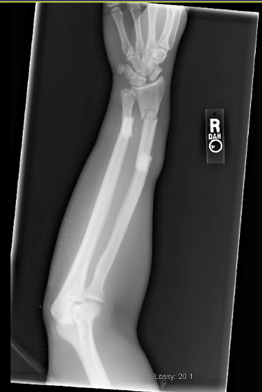
The following case was a high energy trauma victim that was taken to the operating from the emergency department. This is complex fracture involving the forearm. There was a segmental radius fracture with an associated distal ulnar shaft fracture. The technical tips for this surgery are the following:
-The radius was exposed through an extensile volar Henry approach. Start the dissection distally, keeping the FCR and the radial artery retracted medially and extend the interval proximally as needed. Distally it is easier to identify the structures. Keep in mind that in the classic Henry approach the FCR and radial artery are retracted medially. This ensures that the radial artery will be on the medial side when the incision is extended proximally. In the modified Henry approach the FCR is retracted medially and the radial artery laterally. If a modified approach is used then when you extend the incision proximally the radial artery will be crossing the surgical field as you develop the brachialis and FCR interval
-Pay attention to the bow of the radius and make every effort to restore it.
-Convert the three segments to two segments with a use of a lag screw when possible. That facilitates anatomic alignment of the fracture
-Most plates that are not precontoured will need bending to fit the radius.
-Use 3.5mm non locking plates for no articular fractures
-Fixation of the intermediate segment requires two screws for rotational control when one plate is used for fixation of the segmented bone. If two plates are used then you need two screws for each plate applied to the intermediate fragment
-The working length of the plates is more important for biomechanical stability than the total number of screws used
-Partial or complete release of the supinator off the radius proximally may be required. If a complete release is performed leave a cuff of tendon for repair at the end
-Keep the forearm in supination to avoid iatrogenic injury to the posterior interosseous nerve proximally.

Xrays prior and after the fixation of the fracture are shown below.


Friday, August 8, 2014

Distal humerus fractures in the elderly

There is an increasing concern about the distal humerus fractures in the elderly. The concern is about the increasing frequency of this injury in patients with poor bone stock. These patients are usually in their 80s or 90s and pose difficulties in their treatment due to the high levels of activity combined with challenging injuries around the elbow. Open reduction and internal fixation remains the treatment of choice. However, when there is poor bone density fixation can be challenging and the option of total elbow replacement should be entertained. In the years to come orthopaedic surgeons will be doing a lot more total elbow replacements for the reasons explained above. 

The following case is an elderly patient who presented with a distal humerus fracture after a fall. She is physiologically a lot younger than her chronologic age and decided to proceed with operative treatment of the fracture. We prefer, when possible, to do "a triceps on" surgical approach and avoid olecranon osteotomy. The trans-olecranon approach is mandatory when there are multiple intra-articular fragments that require visualization for anatomic reduction and restoration of the smoothness of the articular surface. Xrays before and after surgery are shown and the options of parallel versus biplanar plating are discussed below. In this case two lag screws and orthogonal plating was used for the 3 part distal humerus fracture


Parallel plating
There may be times when it is not possible, because of the specific anatomy of a fracture complex, to place the lateral plate posteriorly, and it has to lie on the crest of the lateral column. In such a scenario, the planes of the two plates will be parallel.
Some surgeons use parallel plating as the preferred method, on the basis that a posterolateral plate permits only short unicortical screws distally, whereas a lateral plate allows the use of longer distal screws.
The choice of method will be determined by the bone quality, the fracture anatomy, the availability of locking plates and individual surgical philosophy.

Biplanar plating
The use of two plates on the distal humerus for C-fractures greatly assists the reconstruction of the triangle of stability. 
When one plate lies on the crest of the medial column, and the other lies on the posterior aspect of the lateral column, which is nonarticular, the two plates lie in planes that are at 90 degrees to each other – perpendicular, or biplanar, plating. 
Such a construct is considered to confer good biomechanical stability in good quality bone, but this has not been proven experimentally.

Wednesday, July 2, 2014

The different faces of shoulder arthritis. Intra-operative pictures

The following case is an elderly woman with many years of pain and crepitus to the shoulder. Her active forward elevation was to 50 degrees with intact rotator cuff. The patient failed conservative treatment and elected to have a total shoulder replacement for her shoulder arthritis. Preoperative, intra-operative and postoperative images are shown below. Most of the time we find loss of articular cartilage and flattening of the humeral head. In this case the arthritic changes were cystic changes to the humeral head and glenoid. Of note the patient has no history of autoimmune disease or inflammatory arthropathy.

Preoperative on the right and postoperative image on the left

Left: Axillary view shows no dislocation. Right: Erosive changes to humeral head and glenoid are seen

Cystic changes to the humeral head were seen intra-operatively

No cartilage was found on the glenoid