Hill Sachs lesion at the posterior superior aspect of the shoulder
Placement of anchor and reimplassage
In French reimplassage means "to fill in". During this procedure the engaging Hill Sachs lesion of the humeral defect is filled in the infraspinatus tendon and the capsule. The Hill Sachs lesion is converted to an extra-articular lesion that does not engage with the anterior glenoid rim providing stability to the shoulder. The procedure was described in 2007 by Wolf et al as an adjunct to the arthroscopic anterior stabilisation procedure of the shoulder in order to address a large engaging Hill-Sach's defect. It is ideally suited to instability patients who have large, engaging Hill-Sachs lesions and soft-tissue Bankart tears. These patients are known to have a higher failure rate after surgery than those with smaller lesions. The results of this technique in this difficult subset of traumatic anterior shoulder instability patients are significantly better (10% recurrence rate) than with an arthroscopic Bankart repair alone (67% recurrence rate). The following case is a patient is his 20s who had 30 dislocations during the past 6 years. His first traumatic anterior shoulder dislocation was a result of a wrestling accident. Due to his young age and no bone loss at the glenoid side we elected to proceed with an arthroscopic Bankart repair combined with a reimplassage procedure due to the large engaging Hill Sachs lesion. Imaging studies and arthroscopy pictures are shown below.
Grashey view
Apical oblique shows no glenoid bone loss
Bankart lesion; view from anterior and posterior portal
Does a preference for "meaningful" work necessitate a lifetime of modest compensation?
Not for those who choose to go into medicine.
That's the finding of a new study from the online salary- and benefits-tracking company PayScale. Using data collected from about 374,000 PayScale site visitors, researchers at PayScale found that doctors tended to have the best combination of high compensation and a positive response to the question "Does your job make the world a better place?" Overall, the group with the best combination of meaning and money is surgeons, 94 percent of whom report finding their work meaningful and whose median compensation was just shy of $300,000. They did, however, also report high levels of stress.
PayScale found that people employed in a group broadly labeled as "community and social service workers"—therapists, clergy, directors of religious programs—were most likely to report that they found their work meaningful, at 84 percent. Clergy, in particular, thought their work was making the world a better place: 97 percent answered in the affirmative.
Unsurprisingly, this group isn't getting rich in a pecuniary sense: Clergy had a median annual pay of $45,500; directors of religious programs made $35,900; marriage and family therapists came in at $47,100. The report noted that jobs in these fields are more likely to be filled by women and that, in general, female-dominated professions were more likely to be high-meaning and low-earning, the lower right-hand corner of the chart below.
PayScale's findings. Size of the circle represents job satisfaction. Click for a larger version. For an interactive version, see here. (PayScale)Of course, many jobs earn little in both cash and fulfillment. Low-paying service jobs—food prep, cashiers, fast-food cooks—were both poorly compensated and not meaningful (the chart's lower-left area). No professions at all fell in the chart's upper-left-hand reaches: highly remunerative and not very meaningful. Those employed in legal services (which includes lawyers in addition to paralegals, clerks, title examiners, etc.) were doing the best of the low-meaning jobs, making $49,000 a year. Lawyers, the top-earners of the bunch, make on average $89,800 and 40 percent said their jobs made the world better. (They too reported high stress.) Financial analysts have the same rate of finding "meaning" in their work as lawyers do, but they make slightly less—$64,900. Over email, PayScale's lead economist Katie Bardaro added that these compensation numbers are national medians, so outliers such as top Wall Street bankers don't push the circle for financial analysts up into the upper-left quadrant.
Of course, not everyone prioritizes finding "meaningful" work, and the report adds that job satisfaction "is not necessarily tied to job meaning." Gaming supervisors—people who run tables in casinos—report high levels of job satisfaction (80 percent) but low meaning (22 percent). OBGYNs, in contrast, don't find their jobs very satisfying but do find them meaningful (they're also paid well for it—more than $200,000).
Lastly, surveys like this give a very broad-brush picture, and any given job can diverge wildly from these reported rates. Do lawyers working in criminal defense feel the same way about their jobs as those working in corporate litigation? Unlikely, but that's not the level of granularity we've got here.
A2 glenoids according to the Walch classification are not difficult to ream because there is no deformity. However, the medial glenoid erosion is best approached with minimal reaming of the glenoid to the subchondral plate. The following case is a patient is her 70s who presented with GH joint arthritis and has failed conservative Rx. She had an intact rotator cuff and active forward shoulder elevation to 100 degrees. The surgical exposure and the preoperative and postoperative XRs are shown below. This case highlights the need of careful reaming to avoid loss of "structural support" on the glenoid side.
Loss of cartilage from humeral head and inferior osteophytes
Exposure of the glenoid
After the drilling of the holes for the central and peripheral pegs of the all polyethylene glenoid component
Implantation of the glenoid component
Placement of 6 #2 Fiberwire sutures for the repair of the subscapularis back to the lesser tuberosity
Implantation of the humeral stem and head
Postopertive Grashey view showing the height and alignment of the prosthesis
The most common reason for revision of the total shoulder replacement is failure of the glenoid component. A few years back the metal backed glenoid components were introduced in an attempt to prolong the longevity of the implant. The idea of component screw fixation to the glenoid bone was challenged by the early failure of the metal backed glenoids. While the main reason of failure of the all polyethylene glenoid components was loosening, the metal backed components fail with several different modes. In our analysis of 4606 arthroplasties performed over a 40 year period it was shown that 1 in 25 all poly glenoid components are revised at 7 years post-op while 1 in 7 metal backed glenoid components needed revision at almost the same fwup time. The study was published in the June 18th issue of the Journal of Bone and Joint Surgery and is authored by Anastasios Papadonikolakis and Frederick A Matsen.
The findings of our study are supported by the recent report of the Australian Total Joint Registry where it is indicated that the early failure of some metal backed designs resulted in removal of specific metal backed designs from the Australian implant market.
In the June 2014 Issue of the American Journal of Sports Medicine the need for acromioplasty during rotator cuff repair is questioned. Please see abstract below. We have pointed out HERE the failure to demonstrate any benefit of acromioplasty in shoulder function.
Arthroscopic Repair of Full-Thickness Rotator Cuff Tears With and Without Acromioplasty
Randomized Prospective Trial With 2-Year Follow-up
†Department of Orthopedic Surgery, Stanford University, Stanford, CA, USA
‡Veterans Administration Palo Alto, Palo Alto, CA, USA
§Florida Orthopedic Institute, Tampa, FL, USA
‖Department of Orthopedic Surgery, Rush University Medical Center, Chicago, IL, USA
¶Department of Orthopedic Surgery, Duke University, Durham, NC, USA
Investigation performed at Rush University Medical Center, Chicago, Illinois, USA
↵* Geoffrey D. Abrams, MD, 3801 Miranda Avenue, Mail Code 112, Palo Alto, CA 94304, USA (e-mail: gabrams@gmail.com).
Presented as a poster at the 39th annual meeting of the AOSSM, Chicago, Illinois, USA, July 2013.
Abstract
Background: Acromioplasty is commonly performed during arthroscopic rotator cuff repair, but its effect on short-term outcomes is debated.
Purpose: To report the short-term clinical outcomes of patients undergoing arthroscopic repair of full-thickness rotator cuff tears with and without acromioplasty.
Study Design: Randomized controlled trial; Level of evidence, 2.
Methods: Patients undergoing arthroscopic repair of full-thickness rotator cuff tears were randomized into acromioplasty or nonacromioplasty groups. The Simple Shoulder Test (SST), American Shoulder and Elbow Surgeons (ASES) score, Constant score, University of California–Los Angeles (UCLA) score, and Short Form–12 (SF-12) health assessment were collected along with physical examination including range of motion and dynamometer strength testing. Intraoperative data including tear size, repair configuration, and concomitant procedures were recorded. Follow-up examination was performed at regular intervals up to 2 years. Preoperative imaging was reviewed to classify the acromial morphologic type, acromial angle, and lateral acromial angulation.
Results: A total of 114 patients were initially enrolled in the study, and 95 (83%; 43 nonacromioplasty, 52 acromioplasty) were available for a minimum 2-year follow-up. There were no significant differences in baseline characteristics, including number of tendons torn, repair configuration, concomitant procedures, and acromion type and angles. Within groups, there was a significant (P < .001) improvement in all functional outcome scores from preoperatively to all follow-up time points, including 2 years, for the nonacromioplasty and acromioplasty groups (ASES score: 55.1-91.5, 48.8-89.0; Constant score: 48.3-75.0, 51.9-78.7, respectively). There were no significant differences in functional outcomes between nonacromioplasty and acromioplasty groups or between subjects with different acromial features at any time point.
Conclusion: The results of this study demonstrate no difference in clinical outcomes after rotator cuff repair with or without acromioplasty at 2 years postoperatively.
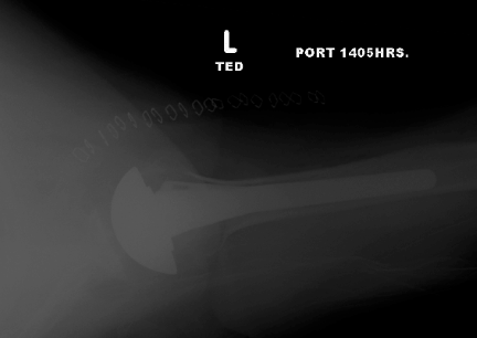
The following case is a 4 year old female who fell off a trampoline. Presented with an angulated proximal humerus fracture. She was treated with a sling. At one year followup the fracture is completely remodelled and there is no visible angulation or fracture line.
There is almost never the need for surgery in this patient population for this type of injury. However, in patients greater than 12 years of age a careful assessment of the degree of angulation and displacement needs to be done .
The indication for surgery are for:
severly displaced fractures in adolescents
<50% apposition or >45° angulation
It is important to remember that the proximal humerus in the pediatric population has:
Three centers of ossification
humeral head appears at 6 mos
greater tuberosity appears at 3 yrs
lesser tuberosity appears at 5 yrs
Secondary ossification centers unite together at age 6-7
Proximal humerus physis closes at 14-17 in girls, 16-18 in boys
80% of humerus growth comes from the proximal physis
highest proximal:distal ratio difference (femur is second with 30:70 proximal:distal ratio)
A 60 y/oF with a failed hemiarthroplasty performed at a University Center presented to our office 1
year after her surgery. Her hemi-arthroplasty was performed for proximal humerus fracture. Her problems at the time of presentation to our office were: a nonunion of tuberosities, loose humeral stem, no subscapularis
function, active forward elevation (FE) to 30 deg, intact deltoid function, anterosuperior
escape with FE of the shoulder
She had no signs of infection at the time of surgery with normal lab values (WBC, CRP, ESR) at the time of presentation. 12 cultures obtained during surgery demonstrated the presence of the p acnes bacterium.
Her problems from this surgery were:
1) Infected loose humeral stem (p acnes)
2) fracture of the humeral stem during revision surgery that required ORIF
3) Neuropraxia of the radial nerve that resolved 10 weeks after her surgery
She was seen 18 months after her operation, she had minimal pain and active forward elevation of the shoulder to 140 degrees. The Xrays are shown below
This preop xray shows that the stem was cemented, there is failure of the tuberosity healing
Anterior dislocation of the prosthesis with motion of the shoulder. Non union of the lesser tuberosity
CT scan shows failure to reproduce the retroversion of the humerus or free rotation of the stem
The failed hemi arthroplasty was revised to a reverse shoulder replacement, the humeral shaft fracture was fixed with 4.5mm DCP plate and eventually healed. Notice the working length of the plate
18 months after surgery and antibiotic treatment for 12 months she has minimal pain. The glenoid component shows no signs of loosening
Her postoperative active shoulder forward elevation is shown below.
Same authors have suggested the use of humeral allograft for the proximal humeral bone loss as seen in this case. There is no high level evidence studies to support the use of allograft. In the setting of a possible low grade p acnes infection the use of allograft would have complicated the infection further. In addition based on the following study the chances of complications after reverse arthroplasty for failed hemi-arthroplasty are high including humeral fracture as in this case.
Reverse total shoulder arthroplasty for the management of failed shoulder arthroplasty with proximal humeral bone loss: is allograft augmentation necessary?
Patients undergoing revision shoulder arthroplasty frequently have deficient proximal humeral bone stock. Proximal humeral allograft has been recommended to augment reverse total shoulder arthroplasty (RTSA) to improve stability and function. This study reports the results of RTSA without proximal humeral allograft in patients with proximal humeral bone loss secondary to failed shoulder arthroplasty.
MATERIALS AND METHODS:
From 2005 to 2008, 251 patients were enrolled in a prospective RTSA cohort study. Significant humeral bone loss was demonstrated in 15 of 56 undergoing revision for failed arthroplasty. Average age was 67 years. Average bone loss measured 38.4 mm (range, 26-72 mm). Patients were followed up for a minimum of 2 years with American Shoulder and Elbow Surgeons (ASES), Subjective Shoulder Value (SSV), Constant Score (CS), and visual analog scale (VAS) pain scores, as well as self-reported satisfaction and radiographs.
RESULTS:
Patients demonstrated significant improvement in mean CS (23.0 to 44.2), ASES (38.2 to 68.3), ASES activities of daily living (7.0 to 15.9), SSV (19.2 to 75.8), and VAS pain (4.6 to 1.6) scores. Thirteen of 15 patients reported satisfaction (87%). Range of motion improved in forward flexion (38.3° to 103.2°) and external rotation (-0.5° to 11.9°). Radiographs demonstrated notching in 3 patients (20%), no humeral subsidence or loosening, and prosthetic fracture of 1 modular humeral stem.
CONCLUSIONS:
Use of RTSA for failed shoulder arthroplasty and deficient humeral bone stock provides a significant clinical benefit without the need for allograft augmentation. Monoblock humeral component use may diminish risk for prosthetic fracture.
The following case is a 55 y/o female with long history of shoulder pain and instability. She was diagnosed with cuff tear arthropathy and has no history of trauma. She has pseudoparalysis of the L shoulder with active FE of the shoulder to 45 degrees and intact deltoid muscle function. Due to pain and inability to maintain her active lifestyle she was treated with a reverse total shoulder replacement and was informed that the chances of glenoid loosening at 10 years postoperatively can be up to 25%.
The picture below demonstrates her active forward elevation
Radiographs below demonstrate a high riding humeral head, superior erosion of the glenoid and loss of glenohumeral joint space.
There are several classification systems for the cuff tear arthropathy however their value for clinical use is limited. A few of those are reported below
Type 1A - Centered stable, Minimal superior migration, C-A arch acetabularization
Type 1B - Centered medialized, Minimal superior migration, medial glenoid erosion, C-A arch acetabularization
Type 2 A - Decentered limited stable, superior translation, superior-medial erosion significant C-A arch acetabularization
Type 2 B - Decentered unstable, anteriorsuperior escape, C-A arch and anterior structures deficient Glenoid erosion in cuff tear arthropathy: Sirveaux Classification Sirveaux et al, JBJS (B), 86: 388-3985, 2004
E0: Humeral head migration without glenoid erosion E1: Concentric glenoid erosion E2: superior glenoid erosion E3: inferior glenoid erosion
Intra-operative pictures are shown below. We found no rotator cuff tendons attached to the humerus with the exception of the subscapularis tendon that was repaired at the end of surgery.
No rotator cuff seen at the time of surgery
45 degree minimal humeral cut at 30 degrees of retroversion
Glenoid exposure. Suction tip placed at the 3 o'clock position
Placement of Guide Tap tilted 15 degrees inferior
After Reaming to the subchondral plate the baseplate was inserted and a 32mm -4 glenosphere was implanted
The humeral component was implanted and sutures were placed on the humerus for repair of the subscapularis tendon.
We prefer to implant the glenosphere first and do a conservative cut on the humeral side. During the trialing process with trail only on the humeral side and sequential reaming of the humeral side and bone resection aims at balancing of the shoulder. This method allows for bone preservation on the humeral side and the chances of intraoperative glenoid fracture are minimized because of no trailing on the glenoid side.
Final Xray is seen below with inferior placement of the glenoid to avoid notching. Minimal humeral bone resection was performed
6 Months after surgery her ROM and active forward elevation has significantly improved as shown below.