Wednesday, April 30, 2014

Is there a role for mini open rotator cuff repair?


Most patients and most orthopaedic surgeons favor the arthroscopic techniques for repair of the rotator cuff tears. Rotator cuff tears can be traumatic or degenerative. It is important to distinguish between the two because traumatic tears that lead to loss of motion or strength are managed with surgery unless the patient has contra-indications for surgery or is of low demand. The healing rate of traumatic tears is higher compared to degenerative tears. In addition, massive rotator cuff tears in the hands of experienced arthroscopists can be treated arthroscopically. I perform 95% of the time arthroscopic rotator cuff surgery due to faster recovery and less chances of infection. However, there is no data as of today to demonstrate that mini open rotator cuff repairs lead to inferior outcomes at 1 year after surgery. In an era of cost effectiveness we have to highlight that an arthroscopic repair of a massive tear (more than two tendons or greater than 5cm in size) may take 2 or 3 hours to be repaired depending on the expertise of the surgeon. In addition, the cost of arthroscopic surgery compared to mini open technique is a lot higher.

The following case is a 52 y/o farmer who had a fall on an outstretched hand and felt a pop in his shoulder. He was unable to forward flex his shoulder after the fall and the MRI demonstrated a two tendon rupture involving the supraspinatus and infraspinatus. The MRI also demonstrates significant retraction of the supraspinatus. Due to his active lifestyle we elected to proceed with repair within 2 weeks from the fall. A mini open technique was used. One year post operatively he has almost full forward flexion to his shoulder and no pain.

Advantages of mini open surgery:
1) Small transverse incision over the anterior inferior aspect of the acromion. The length of the incision is equal to the total length of the arthroscopic small incisions combined together
2) We prefer a Split of the raphe of the deltoid in a longitudinal fashion without detachment of the fibers from the acromion
3) Operative time of 50 minutes. An arthroscopic approach would have taken between 2 to 4 hours for the illustrated case
4) Reduced cost.
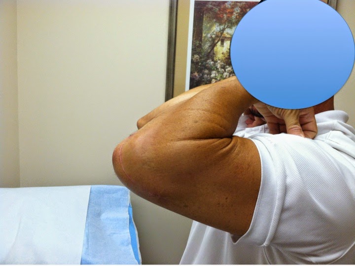
One year after surgery his has almost complete restoration of ROM to the R shoulder. Prior to surgery he has unable to forward flex the shoulder
The arrow indicates the degree of retraction of the supraspinatus after the MRI arthrogram, 

Repaired rotator cuff through deltoid split at the raphe


Clavicle fractures. Reducing the cost of the implant. The use of reconstruction 3.5 mm plates

There is a trend towards operative fixation of shortened and displaced clavicular fractures in high demand young patients.  In the past most fractures of the clavicle were treated with sling immobilization and most orthopaedic surgeons will quote: "The rate of nonunion even for displaced fractures was 0.9%". The debate about these fractures is that it is not important to demonstrate radiographic healing as it was demonstrated in the past. It is also of equal importance to demonstrate that active young patients with malunions returned to their previous job (construction etc) or sports without problems. Studies today show that fragment displacement of more than 100% and shortnening > 2 cm may lead to 30% loss of strength to the shoulder. It is important to let active patients now about the functional limitations if they elect not to have surgery.

Due to the changes in the indications for surgery there is a variety of implants in the market today offering locking screws and "anatomically" contoured plates for fixation of the fracture. In our experience these plates are (1) expesive $$$ (2) The fall into the category "one size fits none". In other words they do not provide what they promise ie fitting to the shape of clavicle.

The following case demonstrates the use of a standard 3.5 reconstruction plate that was contoured at the time of surgery to fit the patient's anatomy. We prefer the use of such a plate if the bone is not osteopenic and there is no need for locking screws. It reduces the cost of surgery and provides easier application without compromise of the reduction (pull of the fragments to the shape of the plate and loss of fixation at the time of screw fixation through the plate)

This a 30 y/o car mechanic who works "under the cars" with the shoulder in forward elevation for 8-10 hours a day. He presented with a shortened and displaced clavicular fracture after an ATV injury. Fixation was performed with a 3.5mm reconstruction plate. He healed without complications and went back to his job at 4 months postoperatively. He was released to full duty at 6 months without restrictions and was advised not to have the plate removed due to the risk of fracture through the screw holes.


Humeral head split in the elderly - Is shoulder prosthetic replacement always necessary?

There is a consensus among orthopaedic surgeons that elderly patients who present with a head split after a proximal humerus fracture will require either reverse shoulder replacement or hemi-arthroplasty. An alternative to surgery is always non operative treatment. Open reduction and internal fixation using a plate should be avoided. The reason lies on the disrupted blood supply to the humeral head that will lead to avascular necrosis, possible failure of fixation and prominence of humeral screws with subsequent erosion to the glenoid cartilage and posttraumatic glenohumeral arthritis. While most cases of humeral head split lead to avascular necrosis of the humeral head there is a small percentage of patients who are not candidates for prosthetic replacement after this severe injury to the proximal humerus. In patients who are frequent fallers as in the case reported below there is a concern of periprosthetic humeral fracture after a new fall. It is common to see periprosthetic fractures after reverse or hemi-arthroplasty if the balance problems of the patient are not addressed or can not be addressed. It is our practice to review the patient's reasons for falling. It is of great value to review medications that the patient is taking along with the concurrent medical conditions that lead to balance problems.

The following case demonstrates a head split after a fall. This is a 77 y/o F who has multiple falls due to balance problems. She lives alone and stays active doing work around the house. Ambulates with a cane and depends on her arms for daily living activities. After explaining risks and benefits of sling immobilization, non operative approach and operative approach (prosthetic replacement vs ORIF) the patient elected to proceed with osteosynthesis. We elected to use an intramedullary fibular graft to support the osteopenic bone of the humeral head and provide better blood flow to the compromised humeral head. 18 months after her surgery she has union of the proximal humerus and active FE to 110 on the injured side (opposite shoulder 160 degrees of FE). She is pleased with the outcome and has not pain. Radiographically there is still a small intra-articular step off. While this case demonstrates the value of the intramedullary fibular grafting we cannot conclude that routine use of this technique will lead to healing of the fracture. A larger study is required to evaluate the effectiveness of this method.
Injury films

Intraop fluroscopic image
18 months postop the fracture has healed.

R side is the operated side. ROM 18 months after surgery.

AO technique:
https://www2.aofoundation.org/wps/portal/!ut/p/c0/04_SB8K8xLLM9MSSzPy8xBz9CP0os3hng7BARydDRwN3QwMDA08zTzdvvxBjIwN_I_2CbEdFADiM_QM!/?redfix_url=1302003581263&implantstype=&segment=Proximal&bone=Humerus&classification=11-C2.3&showPage=redfix&treatment=&method=Open%20reduction%3B%20plate%20fixation

Further reading:
http://boneandjoint.org.uk/highwire/filestream/39970/field_highwire_article_pdf/0/423.full-text.pdf

http://www.ncbi.nlm.nih.gov/pubmed/23823048

Tuesday, April 29, 2014

Pediatric medial epicondyle elbow fractures

The medial epicondyle fractures are common in adolescents and usually nearly 50% of them are associated with an elbow dislocation. It is the third most common pediatric elbow fracture (5-10%) behind supracondylar and lateral condyle fractures. Most commonly they are seen between the ages of 9 and 14 years

The following case illustrates one of our patients who had an ATV injury and dislocated his elbow. He is 12 years old and was initially treated with closed reduction of elbow dislocation. The post reduction films demonstrated a displaced medial epicondyle fracture. The indication for fixation of this fracture is displacement of more than 5 mm. Our experience is that even the mildly displaced fractures with displace over time due to the pull of flexor pronator mass. Ulnar neuropathy is uncommon and was not seen in our case. The fracture was repaired with due 2 K Wires that were removed in the office 4 weeks after the surgery. Medial approach was used and the ulnar nerve was visualized and protected. After fixation intra-operative valgus stress was applied and no elbow instability was found


           - technique considerations:
           - ulnar nerve is identified and protected during exposure;
           - K wires are used in young children, where as screws can be used in adolescents; We prefer K         wires due to the fact that no hardware remains in the bone after removal
           - flexor pronator mass and periosteum are repaired with sutures.


Dislocated elbow

Postreduction films show elbow without dislocationa dn medial epicondyle fracture. Notice the medial epicondyle soft tissue swelling on the image on the Right.

4 weeks postop after fixation of the fracture with two medial K wires and immobilization in the long arm cast.

4 months postop the fracture is healed.


6 weeks after removal of the cast. 

6 weeks after removal of the cast


4 months postop restoration of range of motion


Distal triceps ruptures in athletes - Anabolic steroid use

Distal triceps rupture is a rare injury. It is usually seen in heavy weightlifters and is associated with anabolic steroid use. Lacerations are also common however the most common mechanism is eccentric loading of a contracting triceps. Other local and systemic risk factors associated with rupture include local steroid injection, olecranon bursitis, and hyperparathyroidism although these are uncommon. Initial diagnosis may be difficult because a palpable defect is not always present. Pain and swelling may limit the ability to evaluate strength and elbow range of motion. Although plain radiographs are helpful in ruling out other elbow pathology, MRI is used to confirm the diagnosis, classify the injury, and guide management. If there is an avulsion fracture from the olecranon on lateral xray of the elbow and a palpable triceps defect then expensive MRI imaging is not necessary. MRI is needed to rule out the uncommon muscle tendon junction rupture that is difficult to repair or the muscle rupture. Incomplete tears with active elbow extension against resistance are managed nonsurgically. Surgical repair is indicated in active persons with complete tears and for incomplete tears with concomitant loss of strength. Good to excellent results have been reported with surgical repair, and very good results have been achieved even for chronic tears.

The physiologic chances of the anabolic steroid use to the tendon can explain the susceptibility to rupture. Animal studies have shown that anabolic steroid use leads to:
(1) Increased size and diameter of the tendon without increase in the number of the tendon fibers
(2) Loss of elastic properties of the tendon

The following case is a 35 y.o male body builder who felt a pop in his elbow associated with bruising after a forceful triceps extension exercise with heavy weight. His exam demonstrated loss of elbow extension strength and no palpable defect. No nerve damage was identified. He elected to have his triceps rupture reconstructed. Early repair is critical because after 3-4 weeks the tendon retracts.  A repair is usually recommended within 2-3 weeks from injury to avoid contracture of the tendon. We used a double row technique for restoration of the anatomic footprint of the distal triceps tendon. His postoperative rehab protocol was: elbow immobilized in extension with a brace for 2 weeks then 20 of flexion allowed with the hinged brace every week until full ROM. He had restoration of full ROM and no pain to his arm after the reconstruction. Competitive body building was prohibited due to the risk of re-rupture.
Elbow flexion at 8  weeks postop

Note the avulsed bone fragment from the olecranon. This is typical sign of avulsion of the triceps insertion from the olecranon
Elbow extension at 8 weeks postop

Incision was made lateral to the midline with the patient in a lateral decubitus position. The tear was extending to the muscle tendon junction proximally.

Side to side repair was performed proximally and distally double row repair with 5 anchors

Prior to skin closure the peritenon was repair for restoration of nutrient supply and tendon anatomy.


Further reading: http://www.ncbi.nlm.nih.gov/pubmed/23379659

On the biomechanical properties of the double row repair and restoration of the footprint: http://www.ncbi.nlm.nih.gov/pubmed/20200322 

Monday, April 28, 2014

Pediatric supracondylar humerus fracture.

Most pediatric supracondylar humerus fractures are treated in an operative fashion if there is displacement or angulation. Most of those are extension type fractures and contrary to older beliefs medial pins are not critical for rotational stability of the fracture. There are studies today that demonstrate that larger pins, or two or three lateral pins are biomechanically equally effective compared to combined lateral and medial pinning. Medial pinning should be avoided due to risks of iatrogenic injury to the ulnar nerve. If need to be placed then I would recommend making a small medial elbow incision to visualize the ulnar nerve and the insert the medial pin safely.

Further reading: http://www.ncbi.nlm.nih.gov/pubmed/22706457
http://www.ncbi.nlm.nih.gov/pubmed/22327455

In the following case a Gartland extension type II supracondylar humerus fracture is illustrated. The patient was a 4 year old male that fell of the monkey bars. Was neurovascurarly intact at the time of evaluation. A few technical tips for fixation of these fractures are (1) use a hand table and avoid rotation of the elbow or arm when trying to obtain lateral XRs. Rotation of the arm that may lead to loss of reduction (2) aspirate the hematoma at the olecranon tip prior to reduction  - that facilitates easier reduction (3) Advance the lateral pin just proximal to the fracture line and use it as a joystick to manipulate the distal fragment


Patient regained all ROM after fixation and had no complication


Proximal humerus fracture - dislocation 2

The following case although has the same mechanism of injury as described in the previous post of this blog it was a lot easier to fix and had full restoration of motion at 6 months postoperatively with no pain.

This is a 50 year old  male who works as construction worker. The mechanism of injury was a fall from height. He presented in the ER  where he was diagnosed with R shoulder fracture dislocation. Initially his dislocation was reduced, the shoulder was immobilized in a sling and 2 weeks later he had open reduction and internal fixation of the greater tuberosity fracture using only 2 lag screws.


Proximal humerus fracture - dislocation

The following case is a patient that was seen in the office after a fall from height. He is a 25 year old male who presented with pain to his R shoulder in the  ER and diagnosed with a proximal humerus fracture dislocation. Closed reduction performed by the ER physicians. He seen in our office and reported pain to the R shoulder, denied elbow or hand pain, denied numbness or tingling to the hand.

The preop images demonstrate a significant size greater tuberosity fracture with comminution. Most shoulder surgeons will agree that an anterior traumatic shoulder dislocation as demonstrated in this case is associated with a Hill Sachs lesion and that a fracture of the great tuberosity is rare.

This patient underwent ORIF of the fracture through a deltopectoral approach without detachement of the deltoid. The alternative approach would have been an extensile U shaped incision just lateral to the acromion with a takedown of the deltoid to access the posterosuperior aspect of the humeral head where the fracture is most often located. The disadvantage of this approach is that detachment of the deltoid can cause malfunction of the muscle in addition to higher chances of axillary nerve damage. We selected to use a 3.5 reconstruction plate because it can be contured intraoperatively to match the anatomy of the patient and be positioned in the posterolateral aspect of the humerus where a "typical" locking plate would have been ideal for a lateral side application. In addition, the cost of that plate is significantly less compared to a locking proximal humerus plate.

In this case there were avulsion fractures of the infraspinatus and supraspinatus that required buttressing of these fractures in addition to the repair of the tendon to bone (rotator cuff) with Fiberwire non absorbable sutures.


The glenoid component in total shoulder replacement

Over the course of the past 30 years there have been a lot of different implant designs introduced in the treatment of arthritis of the shoulder. Although most would think that the new implants will have better longevity it seems that this is not true. Based on the largest meta-analysis performed as of today (1) The survivorship of the glenoid component has not changed the past 30 years (data from 1976-2007) (2) Implant design does not affect the longevitity of the prosthesis (13 different implant designs examined) (3) American Shoulder and Elbow Society surgeon membership did not influence the results either

However, the pegged all polythelene glenoid components outperform the keeled in terms of survivorship (see  reference 1) and the metal backed glenoid components fail more often and by different modes compared to all polyethylene glenoid components (in print unpublished data).

Reference 1: http://www.ncbi.nlm.nih.gov/pubmed/24352774

On the modes of failure of the glenoid: http://www.ncbi.nlm.nih.gov/pubmed/18381328

Review on the evolution of the design of the glenoid component: http://reviews.jbjs.org/content/1/2/e2

We prefer to use the all polyethylene pegged cemented glenoid components which allow bone ingrowth at the fenestrations of the central peg. After drilling the hole for the central peg of the glenoid the bone removed is used as bone graft for the central peg.

The radiographs below demonstrate that at 4 months postop there is bony ingrowth at the fenestrations of the central peg and no radiolucent lines.


Radiographically at 10 years 50% of the glenoid components are loose. However the revision rate is low at that point and most of the glenoids "survive" to 10-15 years if the end point is considered to be revision for glenoid failure.