The physiologic chances of the anabolic steroid use to the tendon can explain the susceptibility to rupture. Animal studies have shown that anabolic steroid use leads to:
(1) Increased size and diameter of the tendon without increase in the number of the tendon fibers
(2) Loss of elastic properties of the tendon
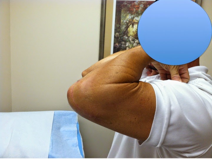
The following case is a 35 y.o male body builder who felt a pop in his elbow associated with bruising after a forceful triceps extension exercise with heavy weight. His exam demonstrated loss of elbow extension strength and no palpable defect. No nerve damage was identified. He elected to have his triceps rupture reconstructed. Early repair is critical because after 3-4 weeks the tendon retracts. A repair is usually recommended within 2-3 weeks from injury to avoid contracture of the tendon. We used a double row technique for restoration of the anatomic footprint of the distal triceps tendon. His postoperative rehab protocol was: elbow immobilized in extension with a brace for 2 weeks then 20 of flexion allowed with the hinged brace every week until full ROM. He had restoration of full ROM and no pain to his arm after the reconstruction. Competitive body building was prohibited due to the risk of re-rupture.
 |
| Elbow flexion at 8 weeks postop |
 |
| Note the avulsed bone fragment from the olecranon. This is typical sign of avulsion of the triceps insertion from the olecranon |
 |
| Elbow extension at 8 weeks postop |
 |
| Incision was made lateral to the midline with the patient in a lateral decubitus position. The tear was extending to the muscle tendon junction proximally. |
 |
| Side to side repair was performed proximally and distally double row repair with 5 anchors |
 |
| Prior to skin closure the peritenon was repair for restoration of nutrient supply and tendon anatomy. |
Further reading: http://www.ncbi.nlm.nih.gov/pubmed/23379659
On the biomechanical properties of the double row repair and restoration of the footprint: http://www.ncbi.nlm.nih.gov/pubmed/20200322