Posterior chondrolabral clefts are associated with glenoid dysplasia and 35% of the time are bilateral according to a study published by the Hospital for Special Surgery (HSS). Although it is difficult to determine whether these lesions are associated with multidirectional, posterior or anterior instability it is recommended to perform examination under anesthesia to evaluate the direction of dislocation or subluxation of the humeral head. The following case is a teenager with a 6 month history of subluxations that were associated with spasms of the anterior shoulder musculature. The patient was treated with 6 months of therapy without improvement of the instability symptoms.
Chondrolabral clefts are located in the posterior inferior labrum, according a HSS study. MRI study studies show that are not normal anatomic variants as there are associated with rounded posterior glenoid rims, glenoid dysplasia and future instability.
Schematic preservation of the rounded posterior glenoid rim
Exam under anesthesia and fluoroscopy demonstrated posterior subluxation of the humeral head on the axillary view with the load and shift test.
Anterior directed force with load and shift test shows no dislocation
Posterior subluxation during load and shift test under anesthesia
MRI demonstrates 1. Round posterior glenoid rim 2. Chondrolabral cleft at the posterior inferior glenoid
Rounded posterior rim of the glenoid and chondrolabral cleft on MRI
Exam under anesthesia demonstrated no dislocation with the load and shift test.
6 o ' clock glenoid
Chondrolabral cleft posterior
Arthroscopic load and shift test demonstrated posterior subluxation of the humeral head (+2 on the load and shift test).
Anterior directed force shows no subluxation during load and shift test
Posterior directed force during load and shift test shows posterior subluxation
Capsulolabral repair using three sutures
One suture anteriorly at the bankart area was placed to complete the capsulolabral repair.
Arthroscopic capsulolabral repair was performed at the area of the cleft. An additional suture for reduction of the capsular volume was placed at the Bankart area where the capsulolabral repair was re-enforced. No anchors were used in this case.
It is speculated that the glenoid dysplasia in combination with the loss of the concavity compression and negative suction pressure leads to instability. Closure of the cleft and shoulder joint capsule reduction seems to be an effective method of addressing the problem.
Tear of the repaired subscapularis tendon after anatomic total shoulder replacement, or hemiarthroplasty remains the main concern that slows down the rehab process. In cases of rupture or stretching of the repaired subscapularis there is external rotational lag or excessive passive external rotation during clinical exam. Radiographically there can be anterior subluxation of the humeral head on axillary view of the shoulder.
I prefer to use 6 sutures for repair of the subscapularis when I re-attach it back to the humerus. The benefit of this approach is that if stretching or rupture occurs occasionally the lower 1/3 of the repaired subscapularis remains attached and thus the tendon does not retract. This is demonstrated in the picture below (C). If the rupture is complete then there is shortening and revision repair back to the lesser tuberosity may required. In such cases soft tissue releases or even augmentation with tendon allograft may be required (D)
Below there is an example of failure of the repaired subscapularis which was was found during surgery to be only stretching of the upper 2/3 of the row border of the tendon. For that reason it was easy to repair it back to the bone. Axillary xray indicates the rupture below.
This axillary view indicates that the humeral head in centered on the glenoid after the revision repair of the subscapularis tendon
This axillary view indicates anterior subluxation, failure of subscapularis
Six sutures passed through the cortical bone of the lesser tuberosity for repair of the subscapularis. Usually the upper 2/3 of the tendon fails at the repair site, or gets stretched and the lower 1/3 remains attached which helps to avoid complete retraction and shortening of the subscapularis tendon after rupture.
The long term outcomes of total shoulder replacement are known. The industry is currently developing hybrid glenoid components, however the all polyethylene glenoid component remains the gold standard in terms of longevity. Metal backed glenoid components have failed to solve the problem of radiolucent lines seen more frequently 5 years after total shoulder replacement. The majority of patients with such a radiographic finding have no clinical adverse event. The following radiographs are 15 years after cemented anatomic total shoulder replacement. The rotator cuff has not failed and the outcome was excellent with active forward elevation to 160 degrees and no pain. There is some polyethylene wear which is expected.
Conservative treatment of the Holstein Lewis humeral shaft fractures remains a very attractive option because there is no risk of iatrogenic radial nerve injury or infection. In addition, the multi-fragmented subgroup of those rare fractures run the risk of non union after operative treatment as there is soft tissue dissection during surgery which disrupts the blood flow to the fracture fragments.
If after application of a Sarmiento brace the fracture shortening is less than 1 cm and the angulation on the AP and lateral plane less than 30 degrees then conservative treatment should be considered.
A case example is shown below that ended up in satisfactory outcome and healing in acceptable position.
Injury film
After application of the Sarmiento brace, AP view
After application of the Sarmiento brace, LAT view
3 months after the fracture is healed
3 months after the fracture is healed
A similar case is illustrated below.
Injury film
Sarmiento application
70 days after the injury the fracture is healed with no angulation of the lateral view
70 days after the injury the fracture is healed with 17 degrees of angulation on the AP view which is within the acceptable limits of malunion
The patient has no function deficits in the ROM of the elbow and is pain free.
Often, there is a concern when inferior subluxation is seen after a proximal humerus fracture. This case below illustrates the problem and how it can be addressed. Most of the time no surgery is required for the "subluxation" as it is not clear what the cause of his phenomenon is. Could be intra-articular blood collection, or hemarthrosis. Could be associated with axillary n neuropraxia which relaxes the deltoid and leads to inferior displacement of the humerus. If no nerve damage and no dislocation is diagnosed, treatment should be in a sling or ORIF depending on other factors and not on subluxation. No surgery for the subluxation is required.
Inferior subluxation
Axillary view shows no dislocation
Position of the humeral head after application of sling
4 weeks post injury the fracture is healed and forward active elevation was to 110 degrees
No dislocation on scapular Y X-ray
3 months post injury the subluxation is resolved and the fracture is healed
Failed rotator cuff surgery is usually a combination of several factors. Occasionally, it is a result of technical error, which in my experience is attributed to "weak repair" or repair under significant tendon tension which leads to progressive failure of the integrity of the repair. Ernest Codman established the principles of successful treatment of rotator cuff tears in the popular monograph "Rupture of the supraspinatus tendon and other lesions in or about the subacromial bursa" that he distributed to the members of American College of Surgeons at the beginning of the 20th century. In his monograph Codman talks about rotator cuff repair surgery in carefully "selected patients". Codman practiced the art of surgery with unparalleled leadership and established several principles for the successful treatment of rotator cuff disease. Today, "patient selection" remains the cornerstone of successful surgery in almost every field of orthopaedic surgery. But what did he mean when he talked about "patient selection" in rotator cuff surgery?
What makes the practice of medicine an art and not a science is the following:
The ability of the surgeon to see the problem within the patient's living environment, function and health status. The ability to understand the needs of the patient, the activity level, the reliability and willingness of the patient to get better and follow the postoperative instructions is a lifelong learning process for the practicing physician. Any underlying health or personality factors that may affect the outcome need to taken into account before offering complex or even simple reconstructive rotator cuff surgery.
The following cases are examples of fitting a treatment plan or surgery to the profile and problem of a particular patient with a complex rotator cuff tear. A torn rotator cuff should never be approached with the only dilemma in mind "to fix or not to fix the torn tendons". This is illustrated below with two case examples.
Case 1:
The following case represents a patient in his 70s who had 2 prior failed rotator cuff repairs. There were "heroic attempts" at repair of the rotator cuff which is indicated in the x-rays below. The number of the metallic anchors indicate the efforts to make the tendon repair successful. A repair is successful if the following factors can be justified as reported by one of the pioneers in shoulder surgery of our era:
1. Good tendon biology (smoking, diabetes or any other nutrition factors may affect healing)
2. Good tendon quality and quantity ( good elasticity and thickness of the tendon, and tension free repair)
3. Absence of glenohumeral arthritis (in the presence of arthritis the repaired rotator cuff will lead to increased contact pressures within the glenohumeral joint and make the pain worse!)
4. Reliable patient who will follow the rehab protocol and protect the repair for at least 3-6 months.
These x-rays above indicate that the blood flow to the footprint of the rotator cuff is compromised, due to the high number of anchors. In addition, the Grashey view indicates a slight superior migration of the humeral head which is a sign of rotator cuff failure. We elected not obtain a preoperative MRI as on exam the abduction and external rotation strength of the shoulder was 4/5 indicative of involvement of the supraspinatus and infraspinatus. The diagnosis of massive rotator cuff tear was clear.
There was no anterosuperior escape and active forward elevation was to 100 degrees without signs of pseudoparalysis. Although there is mild arthritis and the pain level was 9/10 on the VAS scale, it seems that prior to a reverse total shoulder replacement an attempt to improve motion and pain by removing the anchors shoulder be made. Any prominent anchors should be removed and by performing of what Frederick Matsen calls "the smooth and move" procedure there is good chance of establishing a smooth articulation of the humerus to the coracoacromial (CA) arch.
The smoothness of the CA arch is imperative in allowing painless elevation of the shoulder.
Indeed, in this case loose sutures and prominent anchors were seen and removed. The retracted rotator cuff tendon was debrided to stable margins as seen in the pictures below.
Loose sutures at the footprint of the rotator cuff
Humeral head arthritis does not permit rotator cuff repair as such operation increases contact forces in the GH joint and makes the pain worse!
Prominent anchors were removed
Loose or prominent sutures were removed as well.
VIDEO
VIDEO Massive irreparable rotator cuff tear with tendon of poor quality and quantity
The above operation resulted in decrease of the pain from 9/10 to 2/10 and improved the active forward elevation from 100 degrees to 170 degrees. After this surgery there is no need for dedicated rehab, for protection any tendon repair and no retained hardware that can complicate future reconstructive surgery.
Case 2:
The following case represents the same scenario as in Case 1, however the patient is in his 40s, had two previous failed rotator cuff repairs, there is no glenohumeral joint arthritis, there is slight high riding humeral head and there is some tendon left of good thickness that may permit a repair as indicated on the MRI. However, there is shortening of the tendon and a double or single row rotator cuff repair with anchors is not feasible as there will be significant tension in the repair. The two prior failed repairs highlight the need for a tension free repair.
As seen in the pictures below there is no arthritis on xray and the MRI arthrogram shows supraspinatus tendon tear with retraction of 4 cm from the footprint
The arrow indicates the retracted edge of the supraspinatus.
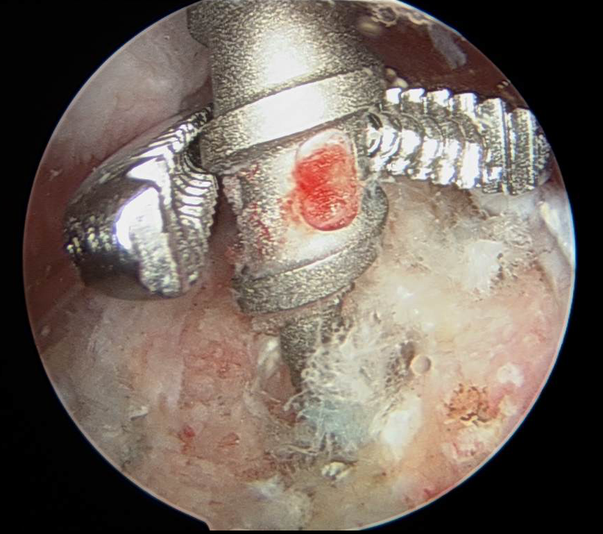
During arthroscopy there was a prominent bioabsorable anchor as seen below. This anchor along with loose suture was removed.
Loose sutures at the fooprint are seen above, viewing from posterior portal with a spinal needle in place
Prominent anchor is seen at the fooprint just anterior to the infraspinatus. Viewing from posterior portal.
A decision was made due to the size and the shape of the tear to proceed with margin convergence which decreased the size of the tear as shown in the drawing below. There was a tendon split at the infraspinatus supraspinatus margin which was incorporated into the margin convergence (video) :
This technique limits superior migration of the humerus during active forward elevation and provides "coverage" of the humeral head with healthy tendon. Biomechanically, when such a tendon to tendon repair heals - which is easier to heal compared to a tendon to bone repair - the forces responsible for humeral head rotation and depression applied by the rotator cuff are partially restored in a suspension bridge configuration as seen below:
Video 1: Size and configuration of the U type tear with an associated substance tear at the infraspinatus supraspinatus margin. The probe is inserted from the anterior portal and the arthroscope from the posterior portal - right shoulder. The area of the prominent anchor that was removed is indicated at the footprint. There is a hole in the rotator cuff footprint of the humerus.
Video 2: Viewing from posterior portal, cannula in the anterior portal, subacromial space, right shoulder. Traction sutures placed in the infraspinatus. Notice the elasticity of the tendon when traction is applied to it. This large size tear cannot be repaired to the footprint by repairing the edges of the tendons back to the fooprint in a "classic" double or single row configuration. In addition, notice the bone loss at the fooprint after the removal of the failed/prominent anchor from prior failed rotator cuff repair.
Video 3: Viewing posterior portal, cannula in the anterior portal, subacromial space, right shoulder. The margin convergence is completed with three non absorbable sutures and nearly 90% of the footprint of the rotator cuff is covered without the need of anchors, tendon transfer or patch.
This case illustrates a successful treatment of a complex shoulder problem. Prior to the introduction of the reverse total shoulder replacement, the operative treatment would have been impossible or very challenging
Rotator cuff arthropathy with glenoid medial erosion and retroversion.