-The radius was exposed through an extensile volar Henry approach. Start the dissection distally, keeping the FCR and the radial artery retracted medially and extend the interval proximally as needed. Distally it is easier to identify the structures. Keep in mind that in the classic Henry approach the FCR and radial artery are retracted medially. This ensures that the radial artery will be on the medial side when the incision is extended proximally. In the modified Henry approach the FCR is retracted medially and the radial artery laterally. If a modified approach is used then when you extend the incision proximally the radial artery will be crossing the surgical field as you develop the brachialis and FCR interval
-Pay attention to the bow of the radius and make every effort to restore it.
-Convert the three segments to two segments with a use of a lag screw when possible. That facilitates anatomic alignment of the fracture
-Most plates that are not precontoured will need bending to fit the radius.
-Use 3.5mm non locking plates for no articular fractures
-Fixation of the intermediate segment requires two screws for rotational control when one plate is used for fixation of the segmented bone. If two plates are used then you need two screws for each plate applied to the intermediate fragment
-The working length of the plates is more important for biomechanical stability than the total number of screws used
-Partial or complete release of the supinator off the radius proximally may be required. If a complete release is performed leave a cuff of tendon for repair at the end
-Keep the forearm in supination to avoid iatrogenic injury to the posterior interosseous nerve proximally.
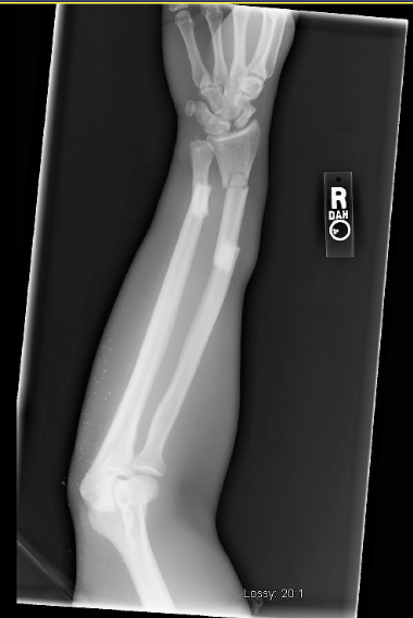
Xrays prior and after the fixation of the fracture are shown below.